Magnesium »
PDB 1g87-1gpm »
1g97 »
Magnesium in PDB 1g97: S.Pneumoniae Glmu Complexed with Udp-N-Acetylglucosamine and MG2+
Enzymatic activity of S.Pneumoniae Glmu Complexed with Udp-N-Acetylglucosamine and MG2+
All present enzymatic activity of S.Pneumoniae Glmu Complexed with Udp-N-Acetylglucosamine and MG2+:
2.7.7.23;
2.7.7.23;
Protein crystallography data
The structure of S.Pneumoniae Glmu Complexed with Udp-N-Acetylglucosamine and MG2+, PDB code: 1g97
was solved by
D.Kostrewa,
A.D'arcy,
M.Kamber,
with X-Ray Crystallography technique. A brief refinement statistics is given in the table below:
| Resolution Low / High (Å) | 30.00 / 1.96 |
| Space group | H 3 2 |
| Cell size a, b, c (Å), α, β, γ (°) | 93.660, 93.660, 275.900, 90.00, 90.00, 120.00 |
| R / Rfree (%) | 18.8 / 22.1 |
Other elements in 1g97:
The structure of S.Pneumoniae Glmu Complexed with Udp-N-Acetylglucosamine and MG2+ also contains other interesting chemical elements:
| Sodium | (Na) | 1 atom |
Magnesium Binding Sites:
The binding sites of Magnesium atom in the S.Pneumoniae Glmu Complexed with Udp-N-Acetylglucosamine and MG2+
(pdb code 1g97). This binding sites where shown within
5.0 Angstroms radius around Magnesium atom.
In total only one binding site of Magnesium was determined in the S.Pneumoniae Glmu Complexed with Udp-N-Acetylglucosamine and MG2+, PDB code: 1g97:
In total only one binding site of Magnesium was determined in the S.Pneumoniae Glmu Complexed with Udp-N-Acetylglucosamine and MG2+, PDB code: 1g97:
Magnesium binding site 1 out of 1 in 1g97
Go back to
Magnesium binding site 1 out
of 1 in the S.Pneumoniae Glmu Complexed with Udp-N-Acetylglucosamine and MG2+
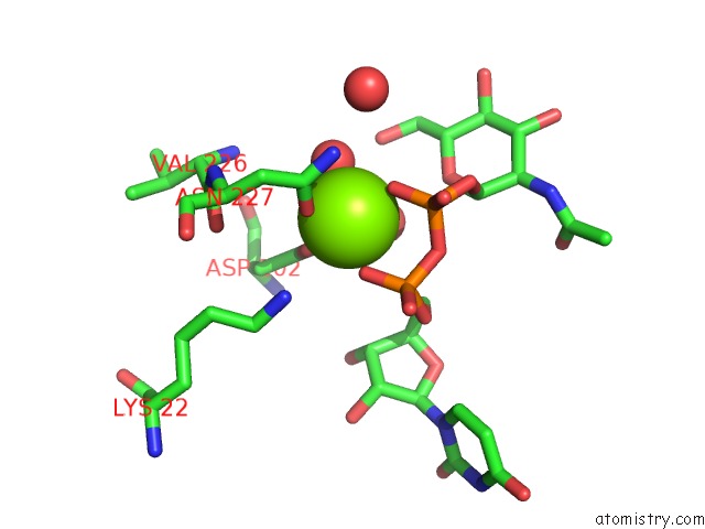
Mono view
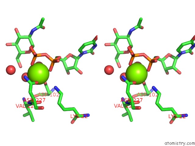
Stereo pair view
Mono view
Stereo pair view
A full contact list of Magnesium with other atoms in the Mg binding
site number 1 of S.Pneumoniae Glmu Complexed with Udp-N-Acetylglucosamine and MG2+ within 5.0Å range:
|
Reference:
D.Kostrewa,
A.D'arcy,
B.Takacs,
M.Kamber.
Crystal Structures of Streptococcus Pneumoniae N-Acetylglucosamine-1-Phosphate Uridyltransferase, Glmu, in Apo Form at 2.33 A Resolution and in Complex with Udp-N-Acetylglucosamine and Mg(2+) at 1.96 A Resolution. J.Mol.Biol. V. 305 279 2001.
ISSN: ISSN 0022-2836
PubMed: 11124906
DOI: 10.1006/JMBI.2000.4296
Page generated: Tue Aug 13 03:42:50 2024
ISSN: ISSN 0022-2836
PubMed: 11124906
DOI: 10.1006/JMBI.2000.4296
Last articles
Zn in 9MJ5Zn in 9HNW
Zn in 9G0L
Zn in 9FNE
Zn in 9DZN
Zn in 9E0I
Zn in 9D32
Zn in 9DAK
Zn in 8ZXC
Zn in 8ZUF