Magnesium »
PDB 1g87-1gpm »
1gag »
Magnesium in PDB 1gag: Crystal Structure of the Insulin Receptor Kinase in Complex with A Bisubstrate Inhibitor
Enzymatic activity of Crystal Structure of the Insulin Receptor Kinase in Complex with A Bisubstrate Inhibitor
All present enzymatic activity of Crystal Structure of the Insulin Receptor Kinase in Complex with A Bisubstrate Inhibitor:
2.7.1.112;
2.7.1.112;
Protein crystallography data
The structure of Crystal Structure of the Insulin Receptor Kinase in Complex with A Bisubstrate Inhibitor, PDB code: 1gag
was solved by
K.Parang,
J.H.Till,
A.J.Ablooglu,
R.A.Kohanski,
S.R.Hubbard,
P.A.Cole,
with X-Ray Crystallography technique. A brief refinement statistics is given in the table below:
| Resolution Low / High (Å) | 30.00 / 2.70 |
| Space group | P 32 2 1 |
| Cell size a, b, c (Å), α, β, γ (°) | 66.300, 66.300, 138.100, 90.00, 90.00, 120.00 |
| R / Rfree (%) | 21.4 / 26.9 |
Magnesium Binding Sites:
The binding sites of Magnesium atom in the Crystal Structure of the Insulin Receptor Kinase in Complex with A Bisubstrate Inhibitor
(pdb code 1gag). This binding sites where shown within
5.0 Angstroms radius around Magnesium atom.
In total 2 binding sites of Magnesium where determined in the Crystal Structure of the Insulin Receptor Kinase in Complex with A Bisubstrate Inhibitor, PDB code: 1gag:
Jump to Magnesium binding site number: 1; 2;
In total 2 binding sites of Magnesium where determined in the Crystal Structure of the Insulin Receptor Kinase in Complex with A Bisubstrate Inhibitor, PDB code: 1gag:
Jump to Magnesium binding site number: 1; 2;
Magnesium binding site 1 out of 2 in 1gag
Go back to
Magnesium binding site 1 out
of 2 in the Crystal Structure of the Insulin Receptor Kinase in Complex with A Bisubstrate Inhibitor
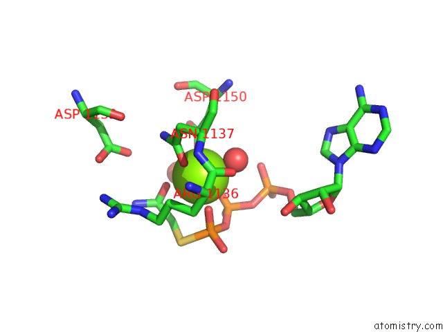
Mono view
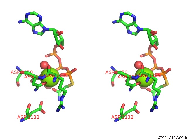
Stereo pair view
Mono view
Stereo pair view
A full contact list of Magnesium with other atoms in the Mg binding
site number 1 of Crystal Structure of the Insulin Receptor Kinase in Complex with A Bisubstrate Inhibitor within 5.0Å range:
|
Magnesium binding site 2 out of 2 in 1gag
Go back to
Magnesium binding site 2 out
of 2 in the Crystal Structure of the Insulin Receptor Kinase in Complex with A Bisubstrate Inhibitor
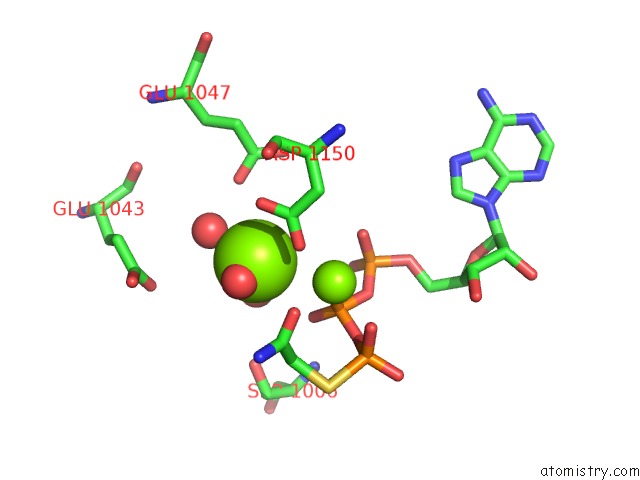
Mono view

Stereo pair view
Mono view
Stereo pair view
A full contact list of Magnesium with other atoms in the Mg binding
site number 2 of Crystal Structure of the Insulin Receptor Kinase in Complex with A Bisubstrate Inhibitor within 5.0Å range:
|
Reference:
K.Parang,
J.H.Till,
A.J.Ablooglu,
R.A.Kohanski,
S.R.Hubbard,
P.A.Cole.
Mechanism-Based Design of A Protein Kinase Inhibitor. Nat.Struct.Biol. V. 8 37 2001.
ISSN: ISSN 1072-8368
PubMed: 11135668
DOI: 10.1038/83028
Page generated: Tue Aug 13 03:43:28 2024
ISSN: ISSN 1072-8368
PubMed: 11135668
DOI: 10.1038/83028
Last articles
Ca in 5T5ICa in 5T57
Ca in 5T4N
Ca in 5T52
Ca in 5T4M
Ca in 5T50
Ca in 5T4Z
Ca in 5T0X
Ca in 5T3H
Ca in 5T2N