Magnesium »
PDB 1g87-1gpm »
1geg »
Magnesium in PDB 1geg: Cryatal Structure Analysis of Meso-2,3-Butanediol Dehydrogenase
Enzymatic activity of Cryatal Structure Analysis of Meso-2,3-Butanediol Dehydrogenase
All present enzymatic activity of Cryatal Structure Analysis of Meso-2,3-Butanediol Dehydrogenase:
1.1.1.5;
1.1.1.5;
Protein crystallography data
The structure of Cryatal Structure Analysis of Meso-2,3-Butanediol Dehydrogenase, PDB code: 1geg
was solved by
M.Otagiri,
G.Kurisu,
S.Ui,
M.Kusunoki,
with X-Ray Crystallography technique. A brief refinement statistics is given in the table below:
| Resolution Low / High (Å) | 40.00 / 1.70 |
| Space group | P 1 21 1 |
| Cell size a, b, c (Å), α, β, γ (°) | 69.160, 109.780, 127.280, 90.00, 102.29, 90.00 |
| R / Rfree (%) | 19.3 / 20.9 |
Magnesium Binding Sites:
The binding sites of Magnesium atom in the Cryatal Structure Analysis of Meso-2,3-Butanediol Dehydrogenase
(pdb code 1geg). This binding sites where shown within
5.0 Angstroms radius around Magnesium atom.
In total 4 binding sites of Magnesium where determined in the Cryatal Structure Analysis of Meso-2,3-Butanediol Dehydrogenase, PDB code: 1geg:
Jump to Magnesium binding site number: 1; 2; 3; 4;
In total 4 binding sites of Magnesium where determined in the Cryatal Structure Analysis of Meso-2,3-Butanediol Dehydrogenase, PDB code: 1geg:
Jump to Magnesium binding site number: 1; 2; 3; 4;
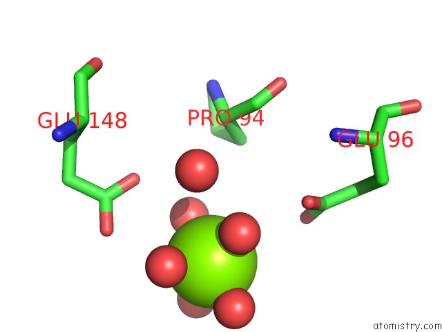
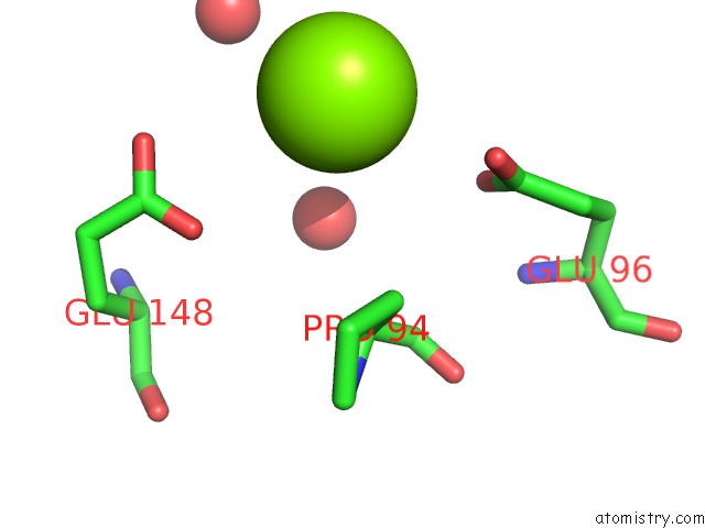
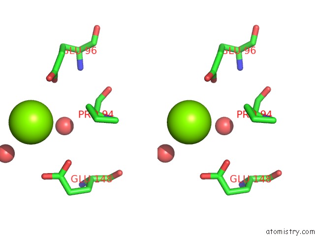
Magnesium binding site 1 out of 4 in 1geg
Go back to
Magnesium binding site 1 out
of 4 in the Cryatal Structure Analysis of Meso-2,3-Butanediol Dehydrogenase
Mono view
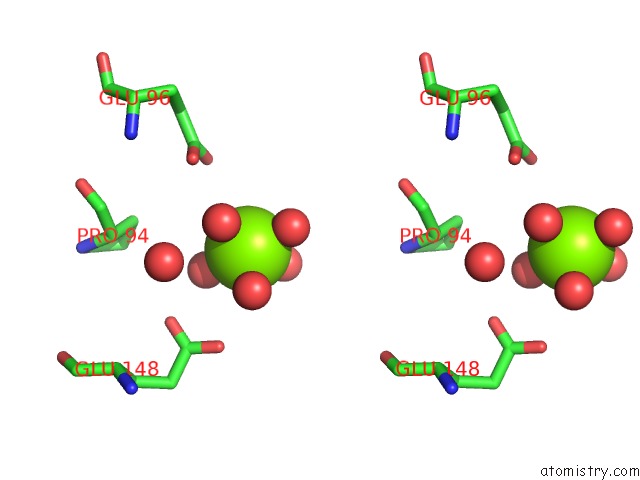
Stereo pair view
Mono view
Stereo pair view
A full contact list of Magnesium with other atoms in the Mg binding
site number 1 of Cryatal Structure Analysis of Meso-2,3-Butanediol Dehydrogenase within 5.0Å range:
|
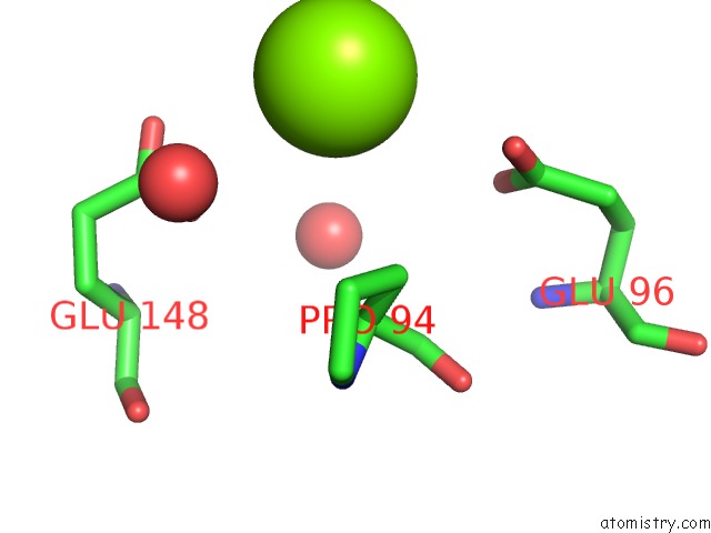
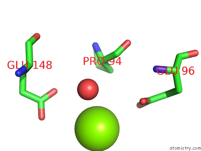
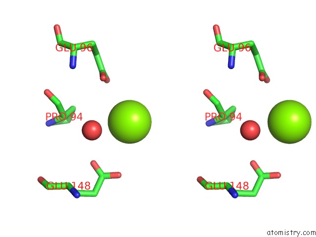
Magnesium binding site 2 out of 4 in 1geg
Go back to
Magnesium binding site 2 out
of 4 in the Cryatal Structure Analysis of Meso-2,3-Butanediol Dehydrogenase
Mono view
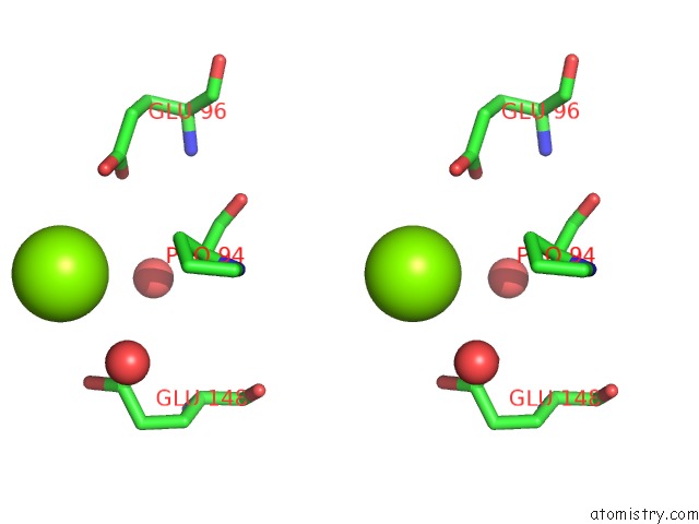
Stereo pair view
Mono view
Stereo pair view
A full contact list of Magnesium with other atoms in the Mg binding
site number 2 of Cryatal Structure Analysis of Meso-2,3-Butanediol Dehydrogenase within 5.0Å range:
|
Magnesium binding site 3 out of 4 in 1geg
Go back to
Magnesium binding site 3 out
of 4 in the Cryatal Structure Analysis of Meso-2,3-Butanediol Dehydrogenase
Mono view
Stereo pair view
Mono view
Stereo pair view
A full contact list of Magnesium with other atoms in the Mg binding
site number 3 of Cryatal Structure Analysis of Meso-2,3-Butanediol Dehydrogenase within 5.0Å range:
|
Magnesium binding site 4 out of 4 in 1geg
Go back to
Magnesium binding site 4 out
of 4 in the Cryatal Structure Analysis of Meso-2,3-Butanediol Dehydrogenase
Mono view
Stereo pair view
Mono view
Stereo pair view
A full contact list of Magnesium with other atoms in the Mg binding
site number 4 of Cryatal Structure Analysis of Meso-2,3-Butanediol Dehydrogenase within 5.0Å range:
|
Reference:
M.Otagiri,
G.Kurisu,
S.Ui,
Y.Takusagawa,
M.Ohkuma,
T.Kudo,
M.Kusunoki.
Crystal Structure of Meso-2,3-Butanediol Dehydrogenase in A Complex with Nad+ and Inhibitor Mercaptoethanol at 1.7 A Resolution For Understanding of Chiral Substrate Recognition Mechanisms. J.Biochem. V. 129 205 2001.
ISSN: ISSN 0021-924X
PubMed: 11173520
DOI: 10.1093/OXFORDJOURNALS.JBCHEM.A002845
Page generated: Tue Aug 13 03:43:49 2024
ISSN: ISSN 0021-924X
PubMed: 11173520
DOI: 10.1093/OXFORDJOURNALS.JBCHEM.A002845
Last articles
Cl in 7YCECl in 7YBX
Cl in 7YAX
Cl in 7YC0
Cl in 7YBP
Cl in 7YBO
Cl in 7Y94
Cl in 7Y6I
Cl in 7YBG
Cl in 7Y7Z