Magnesium »
PDB 1g87-1gpm »
1gim »
Magnesium in PDB 1gim: Crystal Structure of Adenylosuccinate Synthetase From Escherichia Coli Complexed with Gdp, Imp, Hadacidin, NO3-, and MG2+. Data Collected at 100K (pH 6.5)
Enzymatic activity of Crystal Structure of Adenylosuccinate Synthetase From Escherichia Coli Complexed with Gdp, Imp, Hadacidin, NO3-, and MG2+. Data Collected at 100K (pH 6.5)
All present enzymatic activity of Crystal Structure of Adenylosuccinate Synthetase From Escherichia Coli Complexed with Gdp, Imp, Hadacidin, NO3-, and MG2+. Data Collected at 100K (pH 6.5):
6.3.4.4;
6.3.4.4;
Protein crystallography data
The structure of Crystal Structure of Adenylosuccinate Synthetase From Escherichia Coli Complexed with Gdp, Imp, Hadacidin, NO3-, and MG2+. Data Collected at 100K (pH 6.5), PDB code: 1gim
was solved by
B.W.Poland,
H.J.Fromm,
R.B.Honzatko,
with X-Ray Crystallography technique. A brief refinement statistics is given in the table below:
| Resolution Low / High (Å) | 5.00 / 2.50 |
| Space group | P 32 2 1 |
| Cell size a, b, c (Å), α, β, γ (°) | 80.820, 80.820, 159.030, 90.00, 90.00, 120.00 |
| R / Rfree (%) | 20.6 / 27.3 |
Magnesium Binding Sites:
The binding sites of Magnesium atom in the Crystal Structure of Adenylosuccinate Synthetase From Escherichia Coli Complexed with Gdp, Imp, Hadacidin, NO3-, and MG2+. Data Collected at 100K (pH 6.5)
(pdb code 1gim). This binding sites where shown within
5.0 Angstroms radius around Magnesium atom.
In total only one binding site of Magnesium was determined in the Crystal Structure of Adenylosuccinate Synthetase From Escherichia Coli Complexed with Gdp, Imp, Hadacidin, NO3-, and MG2+. Data Collected at 100K (pH 6.5), PDB code: 1gim:
In total only one binding site of Magnesium was determined in the Crystal Structure of Adenylosuccinate Synthetase From Escherichia Coli Complexed with Gdp, Imp, Hadacidin, NO3-, and MG2+. Data Collected at 100K (pH 6.5), PDB code: 1gim:
Magnesium binding site 1 out of 1 in 1gim
Go back to
Magnesium binding site 1 out
of 1 in the Crystal Structure of Adenylosuccinate Synthetase From Escherichia Coli Complexed with Gdp, Imp, Hadacidin, NO3-, and MG2+. Data Collected at 100K (pH 6.5)
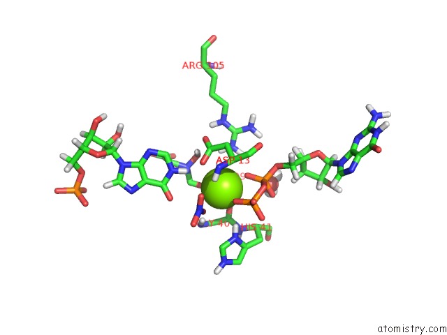
Mono view
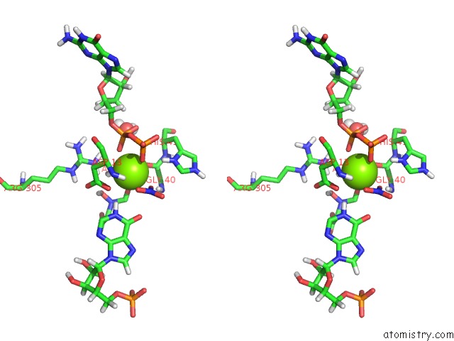
Stereo pair view
Mono view
Stereo pair view
A full contact list of Magnesium with other atoms in the Mg binding
site number 1 of Crystal Structure of Adenylosuccinate Synthetase From Escherichia Coli Complexed with Gdp, Imp, Hadacidin, NO3-, and MG2+. Data Collected at 100K (pH 6.5) within 5.0Å range:
|
Reference:
B.W.Poland,
H.J.Fromm,
R.B.Honzatko.
Crystal Structures of Adenylosuccinate Synthetase From Escherichia Coli Complexed with Gdp, Imp Hadacidin, NO3-, and MG2+. J.Mol.Biol. V. 264 1013 1996.
ISSN: ISSN 0022-2836
PubMed: 9000627
DOI: 10.1006/JMBI.1996.0693
Page generated: Tue Aug 13 03:45:21 2024
ISSN: ISSN 0022-2836
PubMed: 9000627
DOI: 10.1006/JMBI.1996.0693
Last articles
Zn in 9MJ5Zn in 9HNW
Zn in 9G0L
Zn in 9FNE
Zn in 9DZN
Zn in 9E0I
Zn in 9D32
Zn in 9DAK
Zn in 8ZXC
Zn in 8ZUF