Magnesium »
PDB 1g87-1gpm »
1gmi »
Magnesium in PDB 1gmi: Structure of the C2 Domain From Novel Protein Kinase C Epsilon
Protein crystallography data
The structure of Structure of the C2 Domain From Novel Protein Kinase C Epsilon, PDB code: 1gmi
was solved by
W.F.Ochoa,
J.Garcia-Garcia,
I.Fita,
S.Corbalan-Garcia,
N.Verdaguer,
J.C.Gomez-Fernandez,
with X-Ray Crystallography technique. A brief refinement statistics is given in the table below:
| Resolution Low / High (Å) | 20 / 1.7 |
| Space group | P 21 21 21 |
| Cell size a, b, c (Å), α, β, γ (°) | 40.300, 56.500, 60.300, 90.00, 90.00, 90.00 |
| R / Rfree (%) | 23.4 / 26.2 |
Magnesium Binding Sites:
The binding sites of Magnesium atom in the Structure of the C2 Domain From Novel Protein Kinase C Epsilon
(pdb code 1gmi). This binding sites where shown within
5.0 Angstroms radius around Magnesium atom.
In total only one binding site of Magnesium was determined in the Structure of the C2 Domain From Novel Protein Kinase C Epsilon, PDB code: 1gmi:
In total only one binding site of Magnesium was determined in the Structure of the C2 Domain From Novel Protein Kinase C Epsilon, PDB code: 1gmi:
Magnesium binding site 1 out of 1 in 1gmi
Go back to
Magnesium binding site 1 out
of 1 in the Structure of the C2 Domain From Novel Protein Kinase C Epsilon
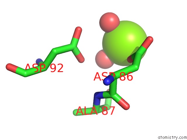
Mono view
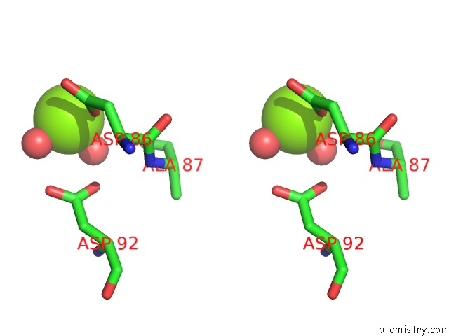
Stereo pair view
Mono view
Stereo pair view
A full contact list of Magnesium with other atoms in the Mg binding
site number 1 of Structure of the C2 Domain From Novel Protein Kinase C Epsilon within 5.0Å range:
|
Reference:
W.F.Ochoa,
J.Garcia-Garcia,
I.Fita,
S.Corbalan-Garcia,
N.Verdaguer,
J.C.Gomez-Fernandez.
Structure of the C2 Domain From Novel Protein Kinase Cepsilon. A Membrane Binding Model For Ca(2+ )-Independent C2 Domains J.Mol.Biol. V. 311 837 2001.
ISSN: ISSN 0022-2836
PubMed: 11518534
DOI: 10.1006/JMBI.2001.4910
Page generated: Tue Aug 13 03:46:59 2024
ISSN: ISSN 0022-2836
PubMed: 11518534
DOI: 10.1006/JMBI.2001.4910
Last articles
F in 4FXYF in 4FXQ
F in 4FZF
F in 4FP1
F in 4FVX
F in 4FV1
F in 4FV9
F in 4FV3
F in 4FS2
F in 4FV0