Magnesium »
PDB 1gq9-1h7q »
1grv »
Magnesium in PDB 1grv: Hypoxanthine Phosphoribosyltransferase From E. Coli
Enzymatic activity of Hypoxanthine Phosphoribosyltransferase From E. Coli
All present enzymatic activity of Hypoxanthine Phosphoribosyltransferase From E. Coli:
2.4.2.8;
2.4.2.8;
Protein crystallography data
The structure of Hypoxanthine Phosphoribosyltransferase From E. Coli, PDB code: 1grv
was solved by
L.W.Guddat,
S.Vos,
J.L.Martin,
D.T.Keough,
J.De Jersey,
with X-Ray Crystallography technique. A brief refinement statistics is given in the table below:
| Resolution Low / High (Å) | 50.0 / 2.90 |
| Space group | P 31 2 1 |
| Cell size a, b, c (Å), α, β, γ (°) | 83.900, 83.900, 169.400, 90.00, 90.00, 120.00 |
| R / Rfree (%) | 21.4 / 23.8 |
Magnesium Binding Sites:
The binding sites of Magnesium atom in the Hypoxanthine Phosphoribosyltransferase From E. Coli
(pdb code 1grv). This binding sites where shown within
5.0 Angstroms radius around Magnesium atom.
In total 2 binding sites of Magnesium where determined in the Hypoxanthine Phosphoribosyltransferase From E. Coli, PDB code: 1grv:
Jump to Magnesium binding site number: 1; 2;
In total 2 binding sites of Magnesium where determined in the Hypoxanthine Phosphoribosyltransferase From E. Coli, PDB code: 1grv:
Jump to Magnesium binding site number: 1; 2;
Magnesium binding site 1 out of 2 in 1grv
Go back to
Magnesium binding site 1 out
of 2 in the Hypoxanthine Phosphoribosyltransferase From E. Coli
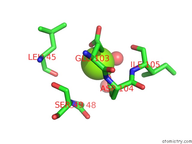
Mono view
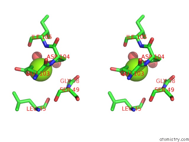
Stereo pair view
Mono view
Stereo pair view
A full contact list of Magnesium with other atoms in the Mg binding
site number 1 of Hypoxanthine Phosphoribosyltransferase From E. Coli within 5.0Å range:
|
Magnesium binding site 2 out of 2 in 1grv
Go back to
Magnesium binding site 2 out
of 2 in the Hypoxanthine Phosphoribosyltransferase From E. Coli
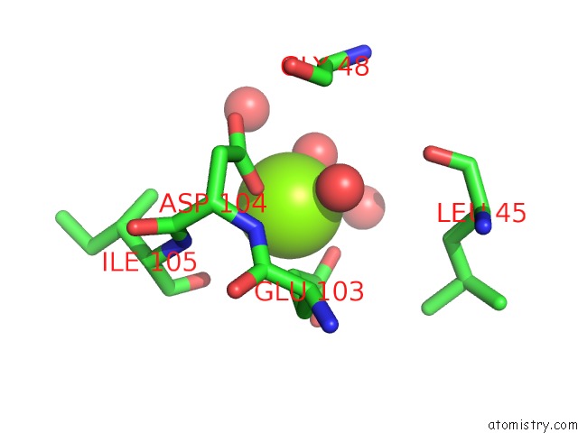
Mono view
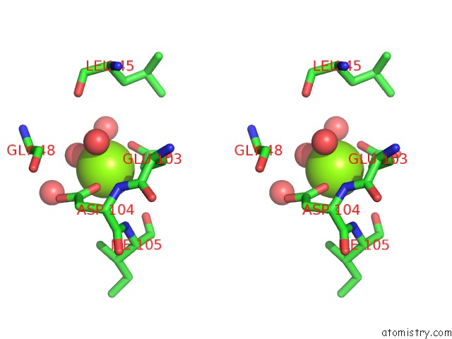
Stereo pair view
Mono view
Stereo pair view
A full contact list of Magnesium with other atoms in the Mg binding
site number 2 of Hypoxanthine Phosphoribosyltransferase From E. Coli within 5.0Å range:
|
Reference:
L.W.Guddat,
S.Vos,
J.L.Martin,
D.T.Keough,
J.De Jersey.
Crystal Structures of Free, Imp-, and Gmp- Bound Escherichia Coli Hypoxanthine Phosphoribosyltransferase Protein Sci. V. 11 1626 2002.
ISSN: ISSN 0961-8368
PubMed: 12070315
DOI: 10.1110/PS.0201002
Page generated: Tue Aug 13 03:50:23 2024
ISSN: ISSN 0961-8368
PubMed: 12070315
DOI: 10.1110/PS.0201002
Last articles
Fe in 2YXOFe in 2YRS
Fe in 2YXC
Fe in 2YNM
Fe in 2YVJ
Fe in 2YP1
Fe in 2YU2
Fe in 2YU1
Fe in 2YQB
Fe in 2YOO