Magnesium »
PDB 1gq9-1h7q »
1gsi »
Magnesium in PDB 1gsi: Crystal Structure of Mycobacterium Tuberculosis Thymidylate Kinase Complexed with Thymidine Monophosphate (Tmp)
Enzymatic activity of Crystal Structure of Mycobacterium Tuberculosis Thymidylate Kinase Complexed with Thymidine Monophosphate (Tmp)
All present enzymatic activity of Crystal Structure of Mycobacterium Tuberculosis Thymidylate Kinase Complexed with Thymidine Monophosphate (Tmp):
2.7.4.9;
2.7.4.9;
Protein crystallography data
The structure of Crystal Structure of Mycobacterium Tuberculosis Thymidylate Kinase Complexed with Thymidine Monophosphate (Tmp), PDB code: 1gsi
was solved by
T.Ursby,
M.Weik,
E.Fioravanti,
M.Delarue,
M.Goeldner,
D.Bourgeois,
with X-Ray Crystallography technique. A brief refinement statistics is given in the table below:
| Resolution Low / High (Å) | 25.19 / 1.6 |
| Space group | P 65 2 2 |
| Cell size a, b, c (Å), α, β, γ (°) | 76.225, 76.225, 134.264, 90.00, 90.00, 120.00 |
| R / Rfree (%) | 19.3 / 21 |
Magnesium Binding Sites:
The binding sites of Magnesium atom in the Crystal Structure of Mycobacterium Tuberculosis Thymidylate Kinase Complexed with Thymidine Monophosphate (Tmp)
(pdb code 1gsi). This binding sites where shown within
5.0 Angstroms radius around Magnesium atom.
In total only one binding site of Magnesium was determined in the Crystal Structure of Mycobacterium Tuberculosis Thymidylate Kinase Complexed with Thymidine Monophosphate (Tmp), PDB code: 1gsi:
In total only one binding site of Magnesium was determined in the Crystal Structure of Mycobacterium Tuberculosis Thymidylate Kinase Complexed with Thymidine Monophosphate (Tmp), PDB code: 1gsi:
Magnesium binding site 1 out of 1 in 1gsi
Go back to
Magnesium binding site 1 out
of 1 in the Crystal Structure of Mycobacterium Tuberculosis Thymidylate Kinase Complexed with Thymidine Monophosphate (Tmp)
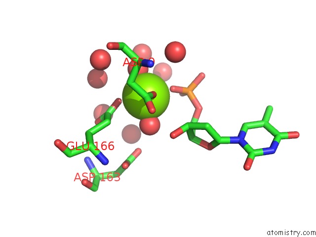
Mono view
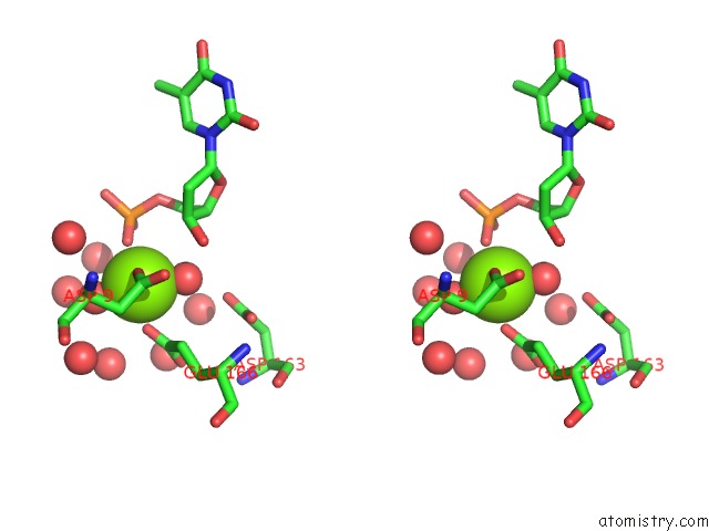
Stereo pair view
Mono view
Stereo pair view
A full contact list of Magnesium with other atoms in the Mg binding
site number 1 of Crystal Structure of Mycobacterium Tuberculosis Thymidylate Kinase Complexed with Thymidine Monophosphate (Tmp) within 5.0Å range:
|
Reference:
T.Ursby,
M.Weik,
E.Fioravanti,
M.Delarue,
M.Goeldner,
D.Bourgeois.
Cryophotolysis of Caged Compounds: A Technique For Trapping Intermediate States in Protein Crystals Acta Crystallogr.,Sect.D V. 58 607 2002.
ISSN: ISSN 0907-4449
PubMed: 11914484
DOI: 10.1107/S0907444902002135
Page generated: Tue Aug 13 03:50:46 2024
ISSN: ISSN 0907-4449
PubMed: 11914484
DOI: 10.1107/S0907444902002135
Last articles
Zn in 9MJ5Zn in 9HNW
Zn in 9G0L
Zn in 9FNE
Zn in 9DZN
Zn in 9E0I
Zn in 9D32
Zn in 9DAK
Zn in 8ZXC
Zn in 8ZUF