Magnesium »
PDB 1gq9-1h7q »
1h1d »
Magnesium in PDB 1h1d: Catechol O-Methyltransferase
Enzymatic activity of Catechol O-Methyltransferase
All present enzymatic activity of Catechol O-Methyltransferase:
2.1.1.6;
2.1.1.6;
Protein crystallography data
The structure of Catechol O-Methyltransferase, PDB code: 1h1d
was solved by
M.Archer,
M.L.Rodrigues,
P.M.Matias,
M.J.Bonifacio,
D.A.Learmonth,
P.Soares-Da-Silva,
M.A.Carrondo,
with X-Ray Crystallography technique. A brief refinement statistics is given in the table below:
| Resolution Low / High (Å) | 25.80 / 2.00 |
| Space group | P 32 2 1 |
| Cell size a, b, c (Å), α, β, γ (°) | 51.490, 51.490, 168.290, 90.00, 90.00, 120.00 |
| R / Rfree (%) | 17.4 / 19.8 |
Other elements in 1h1d:
The structure of Catechol O-Methyltransferase also contains other interesting chemical elements:
| Fluorine | (F) | 3 atoms |
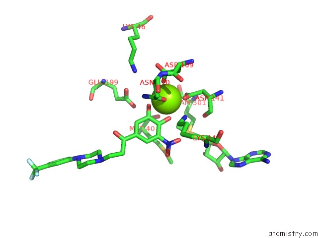
Magnesium Binding Sites:
The binding sites of Magnesium atom in the Catechol O-Methyltransferase
(pdb code 1h1d). This binding sites where shown within
5.0 Angstroms radius around Magnesium atom.
In total only one binding site of Magnesium was determined in the Catechol O-Methyltransferase, PDB code: 1h1d:
In total only one binding site of Magnesium was determined in the Catechol O-Methyltransferase, PDB code: 1h1d:
Magnesium binding site 1 out of 1 in 1h1d
Go back to
Magnesium binding site 1 out
of 1 in the Catechol O-Methyltransferase
Mono view

Stereo pair view
Mono view
Stereo pair view
A full contact list of Magnesium with other atoms in the Mg binding
site number 1 of Catechol O-Methyltransferase within 5.0Å range:
|
Reference:
M.J.Bonifacio,
M.Archer,
M.L.Rodrigues,
P.M.Matias,
D.A.Learmonth,
M.A.Carrondo,
P.Soares-Da-Silva.
Kinetics and Crystal Structure of Catechol-O-Methyltransferase Complex with Co-Substrate and A Novel Inhibitor with Potential Therapeutic Application Mol.Pharmacol. V. 62 795 2002.
ISSN: ISSN 0026-895X
PubMed: 12237326
DOI: 10.1124/MOL.62.4.795
Page generated: Tue Aug 13 03:52:23 2024
ISSN: ISSN 0026-895X
PubMed: 12237326
DOI: 10.1124/MOL.62.4.795
Last articles
Zn in 9MJ5Zn in 9HNW
Zn in 9G0L
Zn in 9FNE
Zn in 9DZN
Zn in 9E0I
Zn in 9D32
Zn in 9DAK
Zn in 8ZXC
Zn in 8ZUF