Magnesium »
PDB 1gq9-1h7q »
1h2a »
Magnesium in PDB 1h2a: Single Crystals of Hydrogenase From Desulfovibrio Vulgaris
Enzymatic activity of Single Crystals of Hydrogenase From Desulfovibrio Vulgaris
All present enzymatic activity of Single Crystals of Hydrogenase From Desulfovibrio Vulgaris:
1.18.99.1;
1.18.99.1;
Protein crystallography data
The structure of Single Crystals of Hydrogenase From Desulfovibrio Vulgaris, PDB code: 1h2a
was solved by
Y.Higuchi,
N.Yasuoka,
with X-Ray Crystallography technique. A brief refinement statistics is given in the table below:
| Resolution Low / High (Å) | 20.00 / 1.80 |
| Space group | P 21 21 21 |
| Cell size a, b, c (Å), α, β, γ (°) | 101.500, 126.500, 66.510, 90.00, 90.00, 90.00 |
| R / Rfree (%) | 22.9 / 27.8 |
Other elements in 1h2a:
The structure of Single Crystals of Hydrogenase From Desulfovibrio Vulgaris also contains other interesting chemical elements:
| Nickel | (Ni) | 1 atom |
| Iron | (Fe) | 12 atoms |
Magnesium Binding Sites:
The binding sites of Magnesium atom in the Single Crystals of Hydrogenase From Desulfovibrio Vulgaris
(pdb code 1h2a). This binding sites where shown within
5.0 Angstroms radius around Magnesium atom.
In total only one binding site of Magnesium was determined in the Single Crystals of Hydrogenase From Desulfovibrio Vulgaris, PDB code: 1h2a:
In total only one binding site of Magnesium was determined in the Single Crystals of Hydrogenase From Desulfovibrio Vulgaris, PDB code: 1h2a:
Magnesium binding site 1 out of 1 in 1h2a
Go back to
Magnesium binding site 1 out
of 1 in the Single Crystals of Hydrogenase From Desulfovibrio Vulgaris
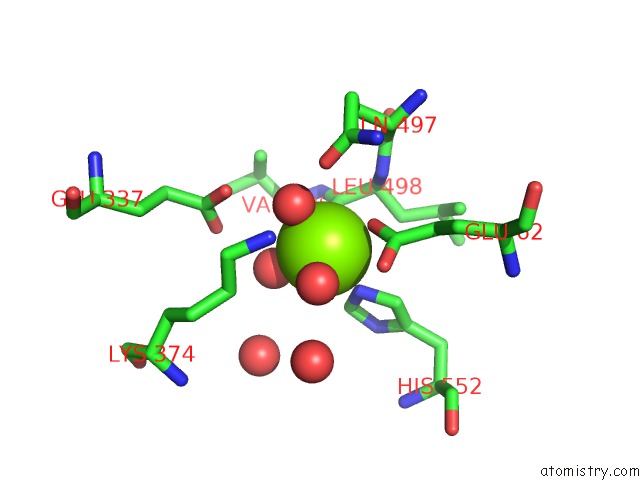
Mono view
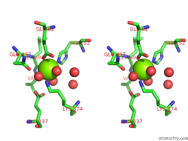
Stereo pair view
Mono view
Stereo pair view
A full contact list of Magnesium with other atoms in the Mg binding
site number 1 of Single Crystals of Hydrogenase From Desulfovibrio Vulgaris within 5.0Å range:
|
Reference:
Y.Higuchi,
T.Yagi,
N.Yasuoka.
Unusual Ligand Structure in Ni-Fe Active Center and An Additional Mg Site in Hydrogenase Revealed By High Resolution X-Ray Structure Analysis. Structure V. 5 1671 1997.
ISSN: ISSN 0969-2126
PubMed: 9438867
DOI: 10.1016/S0969-2126(97)00313-4
Page generated: Tue Aug 13 03:52:41 2024
ISSN: ISSN 0969-2126
PubMed: 9438867
DOI: 10.1016/S0969-2126(97)00313-4
Last articles
Cl in 7SNQCl in 7SNP
Cl in 7SMG
Cl in 7SN0
Cl in 7SF2
Cl in 7SIN
Cl in 7SII
Cl in 7SIL
Cl in 7SHV
Cl in 7SHG