Magnesium »
PDB 1h7u-1hxa »
1hj6 »
Magnesium in PDB 1hj6: Isocitrate Dehydrogenase S113E Mutant Complexed with Isopropylmalate, Nadp+ and Magnesium (Flash-Cooled)
Enzymatic activity of Isocitrate Dehydrogenase S113E Mutant Complexed with Isopropylmalate, Nadp+ and Magnesium (Flash-Cooled)
All present enzymatic activity of Isocitrate Dehydrogenase S113E Mutant Complexed with Isopropylmalate, Nadp+ and Magnesium (Flash-Cooled):
1.1.1.42;
1.1.1.42;
Protein crystallography data
The structure of Isocitrate Dehydrogenase S113E Mutant Complexed with Isopropylmalate, Nadp+ and Magnesium (Flash-Cooled), PDB code: 1hj6
was solved by
S.A.Doyle,
P.T.Beernink,
D.E.Koshland Junior,
with X-Ray Crystallography technique. A brief refinement statistics is given in the table below:
| Resolution Low / High (Å) | 6.0 / 2.0 |
| Space group | P 43 21 2 |
| Cell size a, b, c (Å), α, β, γ (°) | 102.300, 102.300, 150.500, 90.00, 90.00, 90.00 |
| R / Rfree (%) | 20.6 / 24.6 |
Magnesium Binding Sites:
The binding sites of Magnesium atom in the Isocitrate Dehydrogenase S113E Mutant Complexed with Isopropylmalate, Nadp+ and Magnesium (Flash-Cooled)
(pdb code 1hj6). This binding sites where shown within
5.0 Angstroms radius around Magnesium atom.
In total only one binding site of Magnesium was determined in the Isocitrate Dehydrogenase S113E Mutant Complexed with Isopropylmalate, Nadp+ and Magnesium (Flash-Cooled), PDB code: 1hj6:
In total only one binding site of Magnesium was determined in the Isocitrate Dehydrogenase S113E Mutant Complexed with Isopropylmalate, Nadp+ and Magnesium (Flash-Cooled), PDB code: 1hj6:
Magnesium binding site 1 out of 1 in 1hj6
Go back to
Magnesium binding site 1 out
of 1 in the Isocitrate Dehydrogenase S113E Mutant Complexed with Isopropylmalate, Nadp+ and Magnesium (Flash-Cooled)
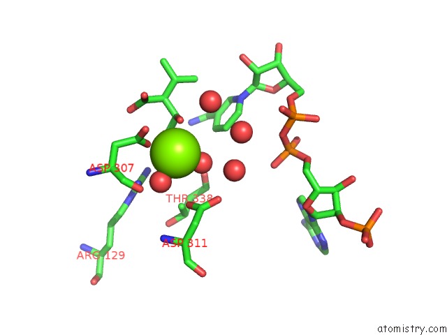
Mono view
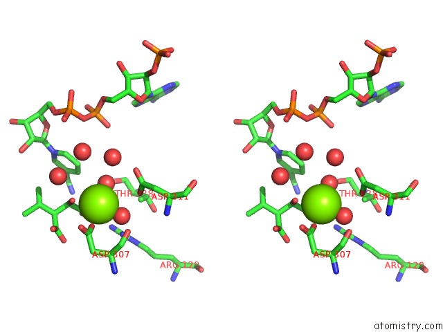
Stereo pair view
Mono view
Stereo pair view
A full contact list of Magnesium with other atoms in the Mg binding
site number 1 of Isocitrate Dehydrogenase S113E Mutant Complexed with Isopropylmalate, Nadp+ and Magnesium (Flash-Cooled) within 5.0Å range:
|
Reference:
S.A.Doyle,
P.T.Beernink,
D.E.Koshland Junior.
Structural Basis For A Change in Substrate Specificity: Crystal Structure of S113E Isocitrate Dehydrogenase in A Complex with Isopropylmalate, MG2+ and Nadp Biochemistry V. 40 4234 2001.
ISSN: ISSN 0006-2960
PubMed: 11284679
DOI: 10.1021/BI002533Q
Page generated: Sat Aug 9 21:29:24 2025
ISSN: ISSN 0006-2960
PubMed: 11284679
DOI: 10.1021/BI002533Q
Last articles
Mg in 3ICZMg in 3ICN
Mg in 3ID8
Mg in 3ICQ
Mg in 3ICE
Mg in 3ICM
Mg in 3ICK
Mg in 3IAQ
Mg in 3IBS
Mg in 3IC4