Magnesium »
PDB 1hxg-1ia0 »
1i44 »
Magnesium in PDB 1i44: Crystallographic Studies of An Activation Loop Mutant of the Insulin Receptor Tyrosine Kinase
Enzymatic activity of Crystallographic Studies of An Activation Loop Mutant of the Insulin Receptor Tyrosine Kinase
All present enzymatic activity of Crystallographic Studies of An Activation Loop Mutant of the Insulin Receptor Tyrosine Kinase:
2.7.1.112;
2.7.1.112;
Protein crystallography data
The structure of Crystallographic Studies of An Activation Loop Mutant of the Insulin Receptor Tyrosine Kinase, PDB code: 1i44
was solved by
J.H.Till,
A.J.Ablooglu,
M.Frankel,
R.A.Kohanski,
S.R.Hubbard,
with X-Ray Crystallography technique. A brief refinement statistics is given in the table below:
| Resolution Low / High (Å) | 30.00 / 2.40 |
| Space group | P 21 21 21 |
| Cell size a, b, c (Å), α, β, γ (°) | 57.857, 69.578, 89.260, 90.00, 90.00, 90.00 |
| R / Rfree (%) | 21 / 26.8 |
Magnesium Binding Sites:
The binding sites of Magnesium atom in the Crystallographic Studies of An Activation Loop Mutant of the Insulin Receptor Tyrosine Kinase
(pdb code 1i44). This binding sites where shown within
5.0 Angstroms radius around Magnesium atom.
In total only one binding site of Magnesium was determined in the Crystallographic Studies of An Activation Loop Mutant of the Insulin Receptor Tyrosine Kinase, PDB code: 1i44:
In total only one binding site of Magnesium was determined in the Crystallographic Studies of An Activation Loop Mutant of the Insulin Receptor Tyrosine Kinase, PDB code: 1i44:
Magnesium binding site 1 out of 1 in 1i44
Go back to
Magnesium binding site 1 out
of 1 in the Crystallographic Studies of An Activation Loop Mutant of the Insulin Receptor Tyrosine Kinase
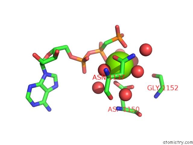
Mono view
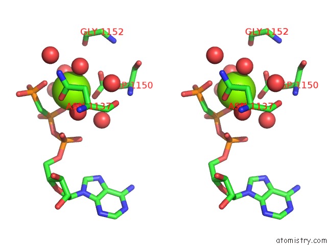
Stereo pair view
Mono view
Stereo pair view
A full contact list of Magnesium with other atoms in the Mg binding
site number 1 of Crystallographic Studies of An Activation Loop Mutant of the Insulin Receptor Tyrosine Kinase within 5.0Å range:
|
Reference:
J.H.Till,
A.J.Ablooglu,
M.Frankel,
S.M.Bishop,
R.A.Kohanski,
S.R.Hubbard.
Crystallographic and Solution Studies of An Activation Loop Mutant of the Insulin Receptor Tyrosine Kinase: Insights Into Kinase Mechanism. J.Biol.Chem. V. 276 10049 2001.
ISSN: ISSN 0021-9258
PubMed: 11124964
DOI: 10.1074/JBC.M010161200
Page generated: Tue Aug 13 04:34:43 2024
ISSN: ISSN 0021-9258
PubMed: 11124964
DOI: 10.1074/JBC.M010161200
Last articles
Zn in 9J0NZn in 9J0O
Zn in 9J0P
Zn in 9FJX
Zn in 9EKB
Zn in 9C0F
Zn in 9CAH
Zn in 9CH0
Zn in 9CH3
Zn in 9CH1