Magnesium »
PDB 1iah-1iru »
1id0 »
Magnesium in PDB 1id0: Crystal Structure of the Nucleotide Bond Conformation of Phoq Kinase Domain
Enzymatic activity of Crystal Structure of the Nucleotide Bond Conformation of Phoq Kinase Domain
All present enzymatic activity of Crystal Structure of the Nucleotide Bond Conformation of Phoq Kinase Domain:
2.7.1.37;
2.7.1.37;
Protein crystallography data
The structure of Crystal Structure of the Nucleotide Bond Conformation of Phoq Kinase Domain, PDB code: 1id0
was solved by
A.Marina,
C.Mott,
A.Auyzenberg,
C.D.Waldburger,
W.A.Hendrickson,
with X-Ray Crystallography technique. A brief refinement statistics is given in the table below:
| Resolution Low / High (Å) | 22.50 / 1.60 |
| Space group | P 21 21 21 |
| Cell size a, b, c (Å), α, β, γ (°) | 43.503, 45.004, 71.764, 90.00, 90.00, 90.00 |
| R / Rfree (%) | 19.9 / 23.3 |
Magnesium Binding Sites:
The binding sites of Magnesium atom in the Crystal Structure of the Nucleotide Bond Conformation of Phoq Kinase Domain
(pdb code 1id0). This binding sites where shown within
5.0 Angstroms radius around Magnesium atom.
In total only one binding site of Magnesium was determined in the Crystal Structure of the Nucleotide Bond Conformation of Phoq Kinase Domain, PDB code: 1id0:
In total only one binding site of Magnesium was determined in the Crystal Structure of the Nucleotide Bond Conformation of Phoq Kinase Domain, PDB code: 1id0:
Magnesium binding site 1 out of 1 in 1id0
Go back to
Magnesium binding site 1 out
of 1 in the Crystal Structure of the Nucleotide Bond Conformation of Phoq Kinase Domain

Mono view
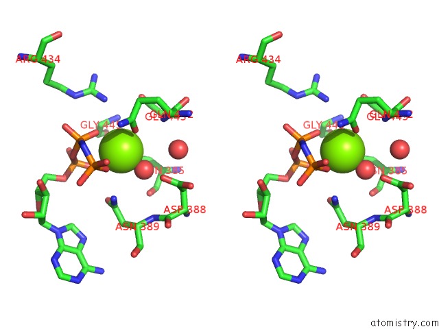
Stereo pair view
Mono view
Stereo pair view
A full contact list of Magnesium with other atoms in the Mg binding
site number 1 of Crystal Structure of the Nucleotide Bond Conformation of Phoq Kinase Domain within 5.0Å range:
|
Reference:
A.Marina,
C.Mott,
A.Auyzenberg,
W.A.Hendrickson,
C.D.Waldburger.
Structural and Mutational Analysis of the Phoq Histidine Kinase Catalytic Domain. Insight Into the Reaction Mechanism. J.Biol.Chem. V. 276 41182 2001.
ISSN: ISSN 0021-9258
PubMed: 11493605
DOI: 10.1074/JBC.M106080200
Page generated: Sat Aug 9 22:14:04 2025
ISSN: ISSN 0021-9258
PubMed: 11493605
DOI: 10.1074/JBC.M106080200
Last articles
Mg in 3T2CMg in 3T2B
Mg in 3T1R
Mg in 3T1O
Mg in 3T0D
Mg in 3T1Q
Mg in 3T12
Mg in 3T1K
Mg in 3T10
Mg in 3T0Z