Magnesium »
PDB 1iah-1iru »
1idc »
Magnesium in PDB 1idc: Isocitrate Dehydrogenase From E.Coli (Mutant K230M), Steady-State Intermediate Complex Determined By Laue Crystallography
Enzymatic activity of Isocitrate Dehydrogenase From E.Coli (Mutant K230M), Steady-State Intermediate Complex Determined By Laue Crystallography
All present enzymatic activity of Isocitrate Dehydrogenase From E.Coli (Mutant K230M), Steady-State Intermediate Complex Determined By Laue Crystallography:
1.1.1.42;
1.1.1.42;
Protein crystallography data
The structure of Isocitrate Dehydrogenase From E.Coli (Mutant K230M), Steady-State Intermediate Complex Determined By Laue Crystallography, PDB code: 1idc
was solved by
J.M.Bolduc,
D.H.Dyer,
W.G.Scott,
P.Singer,
R.M.Sweet,
D.E.Koshland Junior,
B.L.Stoddard,
with X-Ray Crystallography technique. A brief refinement statistics is given in the table below:
| Resolution Low / High (Å) | N/A / 2.50 |
| Space group | P 43 21 2 |
| Cell size a, b, c (Å), α, β, γ (°) | 105.100, 105.100, 150.300, 90.00, 90.00, 90.00 |
| R / Rfree (%) | 16.9 / n/a |
Magnesium Binding Sites:
The binding sites of Magnesium atom in the Isocitrate Dehydrogenase From E.Coli (Mutant K230M), Steady-State Intermediate Complex Determined By Laue Crystallography
(pdb code 1idc). This binding sites where shown within
5.0 Angstroms radius around Magnesium atom.
In total only one binding site of Magnesium was determined in the Isocitrate Dehydrogenase From E.Coli (Mutant K230M), Steady-State Intermediate Complex Determined By Laue Crystallography, PDB code: 1idc:
In total only one binding site of Magnesium was determined in the Isocitrate Dehydrogenase From E.Coli (Mutant K230M), Steady-State Intermediate Complex Determined By Laue Crystallography, PDB code: 1idc:
Magnesium binding site 1 out of 1 in 1idc
Go back to
Magnesium binding site 1 out
of 1 in the Isocitrate Dehydrogenase From E.Coli (Mutant K230M), Steady-State Intermediate Complex Determined By Laue Crystallography
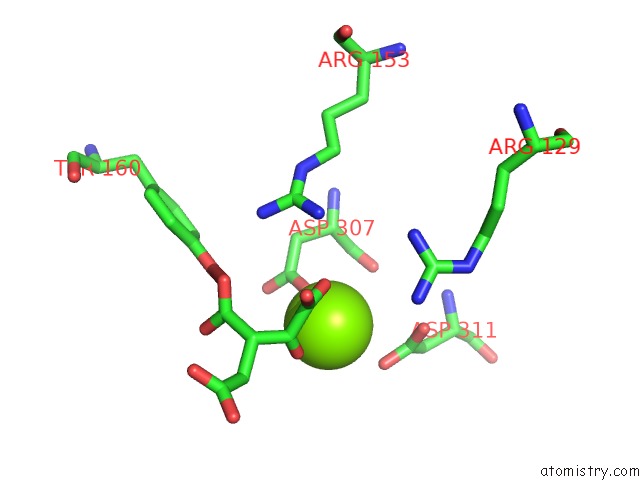
Mono view
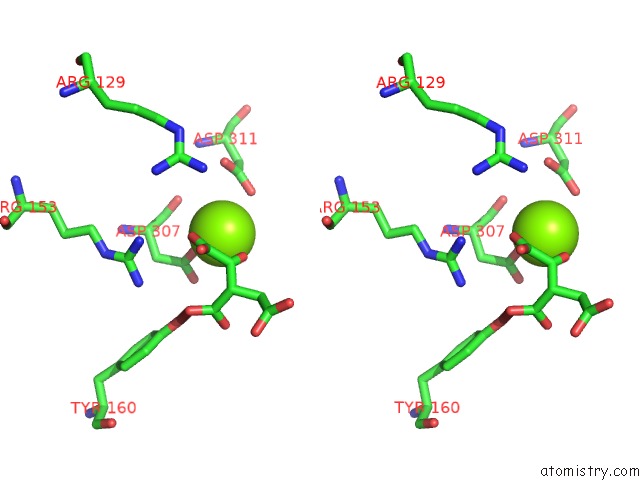
Stereo pair view
Mono view
Stereo pair view
A full contact list of Magnesium with other atoms in the Mg binding
site number 1 of Isocitrate Dehydrogenase From E.Coli (Mutant K230M), Steady-State Intermediate Complex Determined By Laue Crystallography within 5.0Å range:
|
Reference:
J.M.Bolduc,
D.H.Dyer,
W.G.Scott,
P.Singer,
R.M.Sweet,
D.E.Koshland Jr.,
B.L.Stoddard.
Mutagenesis and Laue Structures of Enzyme Intermediates: Isocitrate Dehydrogenase. Science V. 268 1312 1995.
ISSN: ISSN 0036-8075
PubMed: 7761851
Page generated: Sat Aug 9 22:14:20 2025
ISSN: ISSN 0036-8075
PubMed: 7761851
Last articles
Mg in 4CYQMg in 4CYP
Mg in 4CYO
Mg in 4CYJ
Mg in 4CW7
Mg in 4CYN
Mg in 4CYM
Mg in 4CWB
Mg in 4CVN
Mg in 4CT4