Magnesium »
PDB 1it7-1jbz »
1iv3 »
Magnesium in PDB 1iv3: Structure of 2C-Methyl-D-Erythritol-2,4-Cyclodiphosphate Synthase (Bound Form Mg Atoms)
Enzymatic activity of Structure of 2C-Methyl-D-Erythritol-2,4-Cyclodiphosphate Synthase (Bound Form Mg Atoms)
All present enzymatic activity of Structure of 2C-Methyl-D-Erythritol-2,4-Cyclodiphosphate Synthase (Bound Form Mg Atoms):
4.6.1.12;
4.6.1.12;
Protein crystallography data
The structure of Structure of 2C-Methyl-D-Erythritol-2,4-Cyclodiphosphate Synthase (Bound Form Mg Atoms), PDB code: 1iv3
was solved by
H.Kishida,
T.Wada,
S.Unzai,
T.Kuzuyama,
T.Terada,
M.Sirouzu,
S.Yokoyama,
J.R.H.Tame,
S.-Y.Park,
Riken Structuralgenomics/Proteomics Initiative (Rsgi),
with X-Ray Crystallography technique. A brief refinement statistics is given in the table below:
| Resolution Low / High (Å) | 20.00 / 1.52 |
| Space group | P 41 21 2 |
| Cell size a, b, c (Å), α, β, γ (°) | 106.179, 106.179, 148.811, 90.00, 90.00, 90.00 |
| R / Rfree (%) | 20.2 / 25.7 |
Magnesium Binding Sites:
The binding sites of Magnesium atom in the Structure of 2C-Methyl-D-Erythritol-2,4-Cyclodiphosphate Synthase (Bound Form Mg Atoms)
(pdb code 1iv3). This binding sites where shown within
5.0 Angstroms radius around Magnesium atom.
In total 6 binding sites of Magnesium where determined in the Structure of 2C-Methyl-D-Erythritol-2,4-Cyclodiphosphate Synthase (Bound Form Mg Atoms), PDB code: 1iv3:
Jump to Magnesium binding site number: 1; 2; 3; 4; 5; 6;
In total 6 binding sites of Magnesium where determined in the Structure of 2C-Methyl-D-Erythritol-2,4-Cyclodiphosphate Synthase (Bound Form Mg Atoms), PDB code: 1iv3:
Jump to Magnesium binding site number: 1; 2; 3; 4; 5; 6;
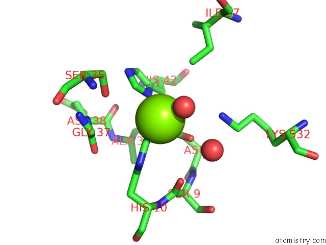
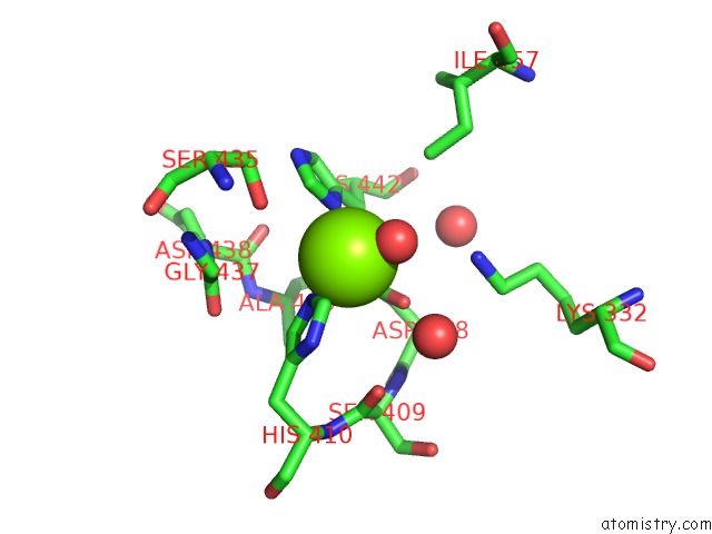
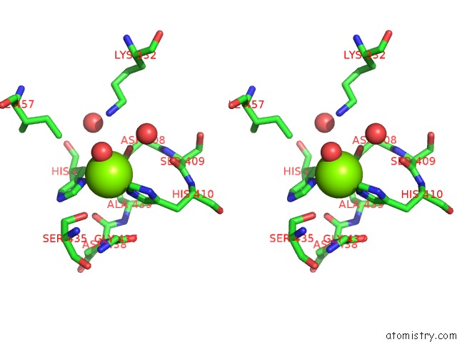
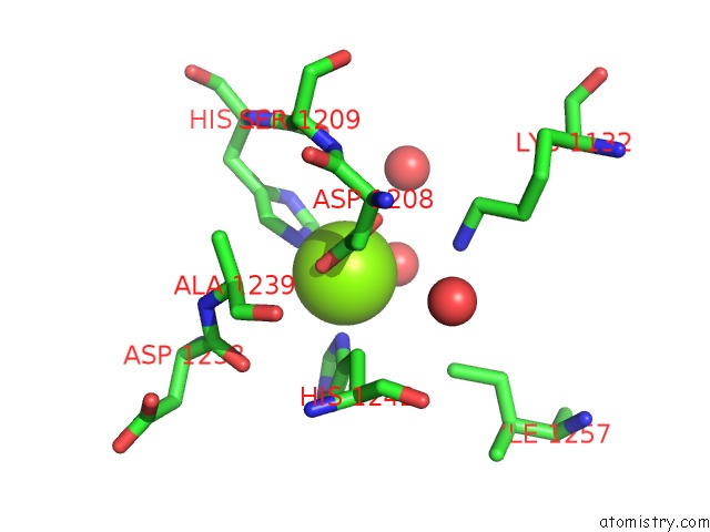
Magnesium binding site 1 out of 6 in 1iv3
Go back to
Magnesium binding site 1 out
of 6 in the Structure of 2C-Methyl-D-Erythritol-2,4-Cyclodiphosphate Synthase (Bound Form Mg Atoms)
Mono view
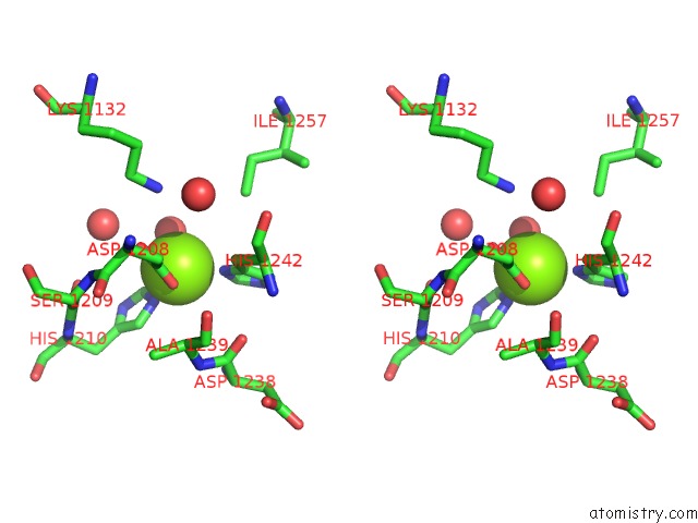
Stereo pair view
Mono view
Stereo pair view
A full contact list of Magnesium with other atoms in the Mg binding
site number 1 of Structure of 2C-Methyl-D-Erythritol-2,4-Cyclodiphosphate Synthase (Bound Form Mg Atoms) within 5.0Å range:
|
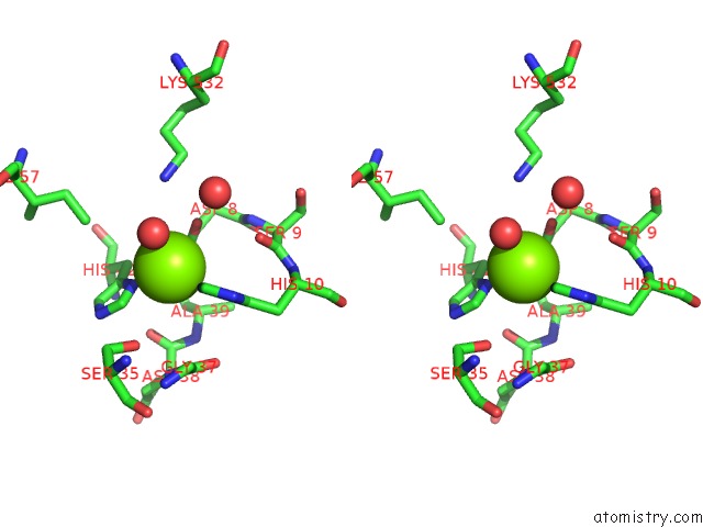
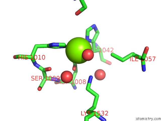
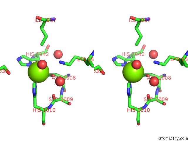
Magnesium binding site 2 out of 6 in 1iv3
Go back to
Magnesium binding site 2 out
of 6 in the Structure of 2C-Methyl-D-Erythritol-2,4-Cyclodiphosphate Synthase (Bound Form Mg Atoms)
Mono view
Stereo pair view
Mono view
Stereo pair view
A full contact list of Magnesium with other atoms in the Mg binding
site number 2 of Structure of 2C-Methyl-D-Erythritol-2,4-Cyclodiphosphate Synthase (Bound Form Mg Atoms) within 5.0Å range:
|
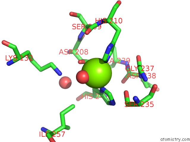
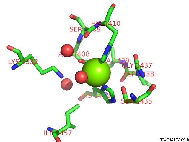
Magnesium binding site 3 out of 6 in 1iv3
Go back to
Magnesium binding site 3 out
of 6 in the Structure of 2C-Methyl-D-Erythritol-2,4-Cyclodiphosphate Synthase (Bound Form Mg Atoms)
Mono view
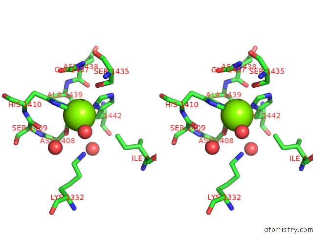
Stereo pair view
Mono view
Stereo pair view
A full contact list of Magnesium with other atoms in the Mg binding
site number 3 of Structure of 2C-Methyl-D-Erythritol-2,4-Cyclodiphosphate Synthase (Bound Form Mg Atoms) within 5.0Å range:
|
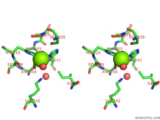
Magnesium binding site 4 out of 6 in 1iv3
Go back to
Magnesium binding site 4 out
of 6 in the Structure of 2C-Methyl-D-Erythritol-2,4-Cyclodiphosphate Synthase (Bound Form Mg Atoms)
Mono view
Stereo pair view
Mono view
Stereo pair view
A full contact list of Magnesium with other atoms in the Mg binding
site number 4 of Structure of 2C-Methyl-D-Erythritol-2,4-Cyclodiphosphate Synthase (Bound Form Mg Atoms) within 5.0Å range:
|
Magnesium binding site 5 out of 6 in 1iv3
Go back to
Magnesium binding site 5 out
of 6 in the Structure of 2C-Methyl-D-Erythritol-2,4-Cyclodiphosphate Synthase (Bound Form Mg Atoms)
Mono view
Stereo pair view
Mono view
Stereo pair view
A full contact list of Magnesium with other atoms in the Mg binding
site number 5 of Structure of 2C-Methyl-D-Erythritol-2,4-Cyclodiphosphate Synthase (Bound Form Mg Atoms) within 5.0Å range:
|
Magnesium binding site 6 out of 6 in 1iv3
Go back to
Magnesium binding site 6 out
of 6 in the Structure of 2C-Methyl-D-Erythritol-2,4-Cyclodiphosphate Synthase (Bound Form Mg Atoms)
Mono view
Stereo pair view
Mono view
Stereo pair view
A full contact list of Magnesium with other atoms in the Mg binding
site number 6 of Structure of 2C-Methyl-D-Erythritol-2,4-Cyclodiphosphate Synthase (Bound Form Mg Atoms) within 5.0Å range:
|
Reference:
H.Kishida,
T.Wada,
S.Unzai,
T.Kuzuyama,
M.Takagi,
T.Terada,
M.Shirouzu,
S.Yokoyama,
J.R.Tame,
S.Y.Park.
Structure and Catalytic Mechanism of 2-C-Methyl-D-Erythritol 2,4-Cyclodiphosphate (Mecdp) Synthase, An Enzyme in the Non-Mevalonate Pathway of Isoprenoid Synthesis. Acta Crystallogr.,Sect.D V. 59 23 2003.
ISSN: ISSN 0907-4449
PubMed: 12499535
DOI: 10.1107/S0907444902017705
Page generated: Sat Aug 9 22:45:21 2025
ISSN: ISSN 0907-4449
PubMed: 12499535
DOI: 10.1107/S0907444902017705
Last articles
Mg in 2WTZMg in 2WTY
Mg in 2WTP
Mg in 2WTO
Mg in 2WSS
Mg in 2WSB
Mg in 2WPD
Mg in 2WOQ
Mg in 2WOJ
Mg in 2WQS