Magnesium »
PDB 1it7-1jbz »
1iyx »
Magnesium in PDB 1iyx: Crystal Structure of Enolase From Enterococcus Hirae
Enzymatic activity of Crystal Structure of Enolase From Enterococcus Hirae
All present enzymatic activity of Crystal Structure of Enolase From Enterococcus Hirae:
4.2.1.11;
4.2.1.11;
Protein crystallography data
The structure of Crystal Structure of Enolase From Enterococcus Hirae, PDB code: 1iyx
was solved by
T.Hosaka,
T.Meguro,
I.Yamato,
Y.Shirakihara,
with X-Ray Crystallography technique. A brief refinement statistics is given in the table below:
| Resolution Low / High (Å) | 20.00 / 2.80 |
| Space group | I 4 |
| Cell size a, b, c (Å), α, β, γ (°) | 153.510, 153.510, 90.660, 90.00, 90.00, 90.00 |
| R / Rfree (%) | 17.5 / 23.9 |
Magnesium Binding Sites:
The binding sites of Magnesium atom in the Crystal Structure of Enolase From Enterococcus Hirae
(pdb code 1iyx). This binding sites where shown within
5.0 Angstroms radius around Magnesium atom.
In total 2 binding sites of Magnesium where determined in the Crystal Structure of Enolase From Enterococcus Hirae, PDB code: 1iyx:
Jump to Magnesium binding site number: 1; 2;
In total 2 binding sites of Magnesium where determined in the Crystal Structure of Enolase From Enterococcus Hirae, PDB code: 1iyx:
Jump to Magnesium binding site number: 1; 2;
Magnesium binding site 1 out of 2 in 1iyx
Go back to
Magnesium binding site 1 out
of 2 in the Crystal Structure of Enolase From Enterococcus Hirae
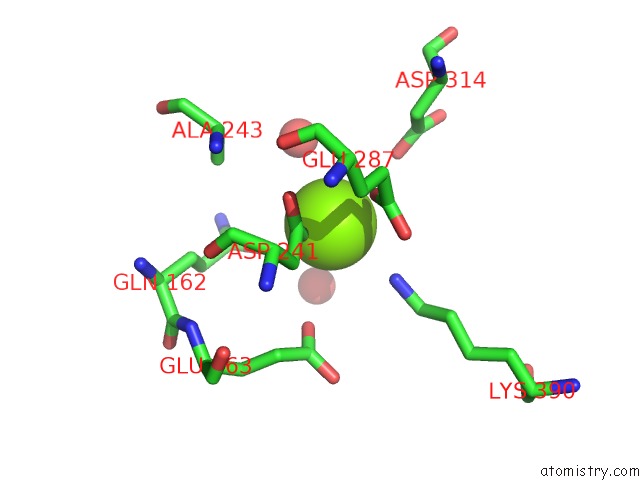
Mono view
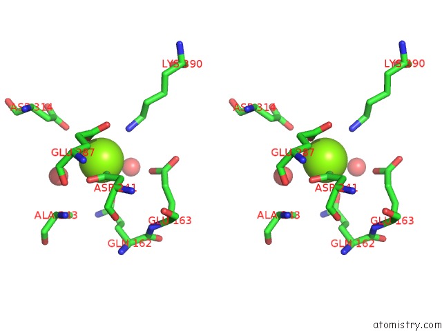
Stereo pair view
Mono view
Stereo pair view
A full contact list of Magnesium with other atoms in the Mg binding
site number 1 of Crystal Structure of Enolase From Enterococcus Hirae within 5.0Å range:
|
Magnesium binding site 2 out of 2 in 1iyx
Go back to
Magnesium binding site 2 out
of 2 in the Crystal Structure of Enolase From Enterococcus Hirae
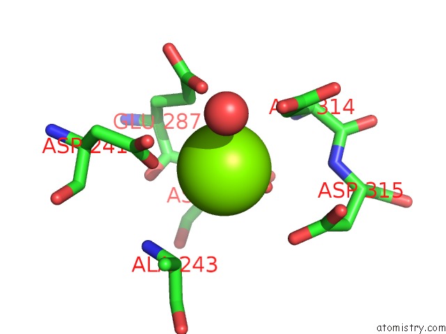
Mono view
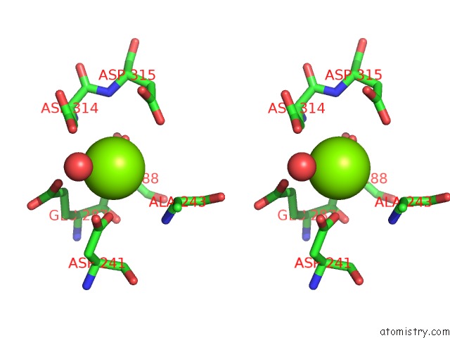
Stereo pair view
Mono view
Stereo pair view
A full contact list of Magnesium with other atoms in the Mg binding
site number 2 of Crystal Structure of Enolase From Enterococcus Hirae within 5.0Å range:
|
Reference:
T.Hosaka,
T.Meguro,
I.Yamato,
Y.Shirakihara.
Crystal Structure of Enterococcus Hirae Enolase at 2.8 A Resolution J.Biochem.(Tokyo) V. 133 817 2003.
ISSN: ISSN 0021-924X
PubMed: 12869539
DOI: 10.1093/JB/MVG104
Page generated: Sat Aug 9 22:47:58 2025
ISSN: ISSN 0021-924X
PubMed: 12869539
DOI: 10.1093/JB/MVG104
Last articles
Mg in 1SO2Mg in 1SO5
Mg in 1SO4
Mg in 1SO3
Mg in 1SNF
Mg in 1SLH
Mg in 1SL2
Mg in 1SL5
Mg in 1SKR
Mg in 1SL0