Magnesium »
PDB 1it7-1jbz »
1j8l »
Magnesium in PDB 1j8l: Molecular and Crystal Structure of D(CGCAAATTMO4CGCG): the Watson-Crick Type N4-Methoxycytidine/Adenosine Base Pair in B-Dna
Protein crystallography data
The structure of Molecular and Crystal Structure of D(CGCAAATTMO4CGCG): the Watson-Crick Type N4-Methoxycytidine/Adenosine Base Pair in B-Dna, PDB code: 1j8l
was solved by
M.T.Hossain,
T.Sunami,
M.Tsunoda,
T.Hikima,
T.Chatake,
Y.Ueno,
A.Matsuda,
A.Takenaka,
with X-Ray Crystallography technique. A brief refinement statistics is given in the table below:
| Resolution Low / High (Å) | 10.00 / 1.60 |
| Space group | P 21 21 21 |
| Cell size a, b, c (Å), α, β, γ (°) | 24.800, 40.300, 65.500, 90.00, 90.00, 90.00 |
| R / Rfree (%) | 21.8 / 25.8 |
Magnesium Binding Sites:
The binding sites of Magnesium atom in the Molecular and Crystal Structure of D(CGCAAATTMO4CGCG): the Watson-Crick Type N4-Methoxycytidine/Adenosine Base Pair in B-Dna
(pdb code 1j8l). This binding sites where shown within
5.0 Angstroms radius around Magnesium atom.
In total only one binding site of Magnesium was determined in the Molecular and Crystal Structure of D(CGCAAATTMO4CGCG): the Watson-Crick Type N4-Methoxycytidine/Adenosine Base Pair in B-Dna, PDB code: 1j8l:
In total only one binding site of Magnesium was determined in the Molecular and Crystal Structure of D(CGCAAATTMO4CGCG): the Watson-Crick Type N4-Methoxycytidine/Adenosine Base Pair in B-Dna, PDB code: 1j8l:
Magnesium binding site 1 out of 1 in 1j8l
Go back to
Magnesium binding site 1 out
of 1 in the Molecular and Crystal Structure of D(CGCAAATTMO4CGCG): the Watson-Crick Type N4-Methoxycytidine/Adenosine Base Pair in B-Dna
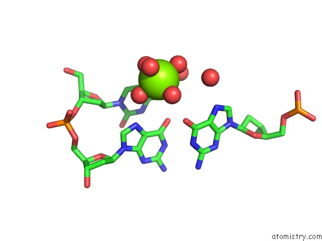
Mono view
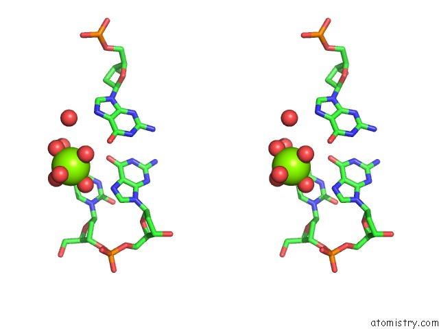
Stereo pair view
Mono view
Stereo pair view
A full contact list of Magnesium with other atoms in the Mg binding
site number 1 of Molecular and Crystal Structure of D(CGCAAATTMO4CGCG): the Watson-Crick Type N4-Methoxycytidine/Adenosine Base Pair in B-Dna within 5.0Å range:
|
Reference:
M.T.Hossain,
T.Sunami,
M.Tsunoda,
T.Hikima,
T.Chatake,
Y.Ueno,
A.Matsuda,
A.Takenaka.
Crystallographic Studies on Damaged Dnas IV. N(4)-Methoxycytosine Shows A Second Face For Watson-Crick Base-Pairing, Leading to Purine Transition Mutagenesis. Nucleic Acids Res. V. 29 3949 2001.
ISSN: ISSN 0305-1048
PubMed: 11574676
Page generated: Tue Aug 13 05:46:28 2024
ISSN: ISSN 0305-1048
PubMed: 11574676
Last articles
Zn in 9J0NZn in 9J0O
Zn in 9J0P
Zn in 9FJX
Zn in 9EKB
Zn in 9C0F
Zn in 9CAH
Zn in 9CH0
Zn in 9CH3
Zn in 9CH1