Magnesium »
PDB 1it7-1jbz »
1jbz »
Magnesium in PDB 1jbz: Crystal Structure Analysis of A Dual-Wavelength Emission Green Fluorescent Protein Variant at High pH
Protein crystallography data
The structure of Crystal Structure Analysis of A Dual-Wavelength Emission Green Fluorescent Protein Variant at High pH, PDB code: 1jbz
was solved by
G.T.Hanson,
T.B.Mcananey,
E.S.Park,
M.E.P.Rendell,
D.K.Yarbrough,
S.Chu,
L.Xi,
S.G.Boxer,
M.H.Montrose,
S.J.Remington,
with X-Ray Crystallography technique. A brief refinement statistics is given in the table below:
| Resolution Low / High (Å) | 19.90 / 1.50 |
| Space group | P 21 21 21 |
| Cell size a, b, c (Å), α, β, γ (°) | 51.200, 62.374, 68.967, 90.00, 90.00, 90.00 |
| R / Rfree (%) | 17.7 / n/a |
Magnesium Binding Sites:
The binding sites of Magnesium atom in the Crystal Structure Analysis of A Dual-Wavelength Emission Green Fluorescent Protein Variant at High pH
(pdb code 1jbz). This binding sites where shown within
5.0 Angstroms radius around Magnesium atom.
In total 2 binding sites of Magnesium where determined in the Crystal Structure Analysis of A Dual-Wavelength Emission Green Fluorescent Protein Variant at High pH, PDB code: 1jbz:
Jump to Magnesium binding site number: 1; 2;
In total 2 binding sites of Magnesium where determined in the Crystal Structure Analysis of A Dual-Wavelength Emission Green Fluorescent Protein Variant at High pH, PDB code: 1jbz:
Jump to Magnesium binding site number: 1; 2;
Magnesium binding site 1 out of 2 in 1jbz
Go back to
Magnesium binding site 1 out
of 2 in the Crystal Structure Analysis of A Dual-Wavelength Emission Green Fluorescent Protein Variant at High pH
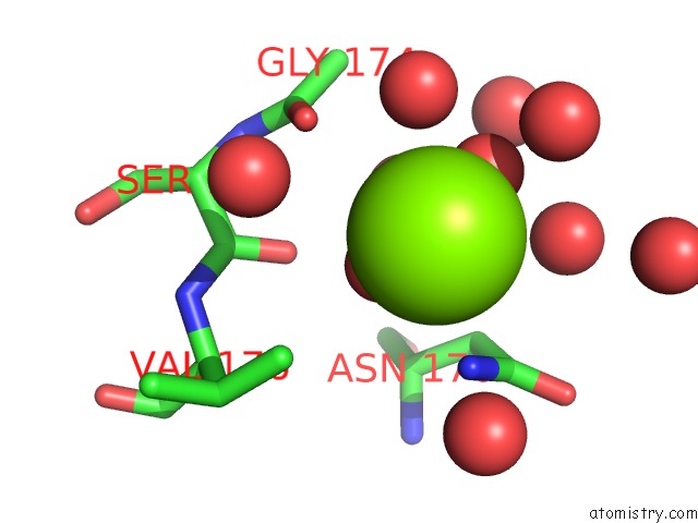
Mono view
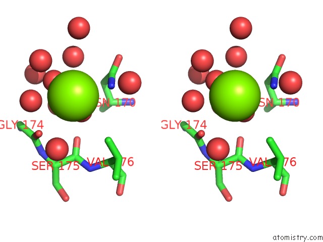
Stereo pair view
Mono view
Stereo pair view
A full contact list of Magnesium with other atoms in the Mg binding
site number 1 of Crystal Structure Analysis of A Dual-Wavelength Emission Green Fluorescent Protein Variant at High pH within 5.0Å range:
|
Magnesium binding site 2 out of 2 in 1jbz
Go back to
Magnesium binding site 2 out
of 2 in the Crystal Structure Analysis of A Dual-Wavelength Emission Green Fluorescent Protein Variant at High pH
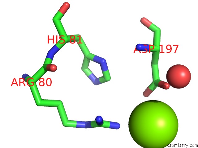
Mono view
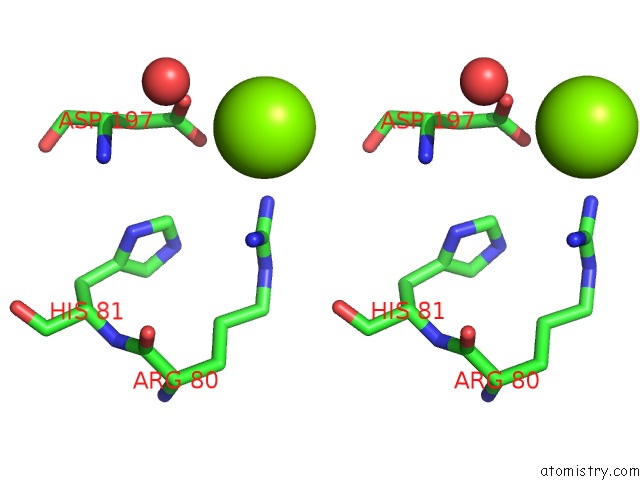
Stereo pair view
Mono view
Stereo pair view
A full contact list of Magnesium with other atoms in the Mg binding
site number 2 of Crystal Structure Analysis of A Dual-Wavelength Emission Green Fluorescent Protein Variant at High pH within 5.0Å range:
|
Reference:
G.T.Hanson,
T.B.Mcananey,
E.S.Park,
M.E.P.Rendell,
D.K.Yarbrough,
S.Chu,
L.Xi,
S.G.Boxer,
M.H.Montrose,
S.J.Remington.
Green Fluorescent Protein Variants As Ratiometric Dual Emission pH Sensors. 1. Structural Characterization and Preliminary Application. Biochemistry V. 41 15477 2002.
ISSN: ISSN 0006-2960
PubMed: 12501176
DOI: 10.1021/BI026609P
Page generated: Sat Aug 9 22:53:42 2025
ISSN: ISSN 0006-2960
PubMed: 12501176
DOI: 10.1021/BI026609P
Last articles
Mg in 4E8PMg in 4E8M
Mg in 4E8G
Mg in 4E84
Mg in 4E89
Mg in 4DV7
Mg in 4E7Z
Mg in 4E6M
Mg in 4E7S
Mg in 4E7P