Magnesium »
PDB 1jcg-1jwy »
1jed »
Magnesium in PDB 1jed: Crystal Structure of Atp Sulfurylase in Complex with Adp
Enzymatic activity of Crystal Structure of Atp Sulfurylase in Complex with Adp
All present enzymatic activity of Crystal Structure of Atp Sulfurylase in Complex with Adp:
2.7.7.4;
2.7.7.4;
Protein crystallography data
The structure of Crystal Structure of Atp Sulfurylase in Complex with Adp, PDB code: 1jed
was solved by
T.C.Ullrich,
R.Huber,
with X-Ray Crystallography technique. A brief refinement statistics is given in the table below:
| Resolution Low / High (Å) | 24.81 / 2.95 |
| Space group | H 3 2 |
| Cell size a, b, c (Å), α, β, γ (°) | 186.135, 186.135, 223.253, 90.00, 90.00, 120.00 |
| R / Rfree (%) | 19.2 / 22.7 |
Other elements in 1jed:
The structure of Crystal Structure of Atp Sulfurylase in Complex with Adp also contains other interesting chemical elements:
| Cadmium | (Cd) | 11 atoms |
| Calcium | (Ca) | 6 atoms |
| Sodium | (Na) | 12 atoms |
Magnesium Binding Sites:
The binding sites of Magnesium atom in the Crystal Structure of Atp Sulfurylase in Complex with Adp
(pdb code 1jed). This binding sites where shown within
5.0 Angstroms radius around Magnesium atom.
In total 4 binding sites of Magnesium where determined in the Crystal Structure of Atp Sulfurylase in Complex with Adp, PDB code: 1jed:
Jump to Magnesium binding site number: 1; 2; 3; 4;
In total 4 binding sites of Magnesium where determined in the Crystal Structure of Atp Sulfurylase in Complex with Adp, PDB code: 1jed:
Jump to Magnesium binding site number: 1; 2; 3; 4;
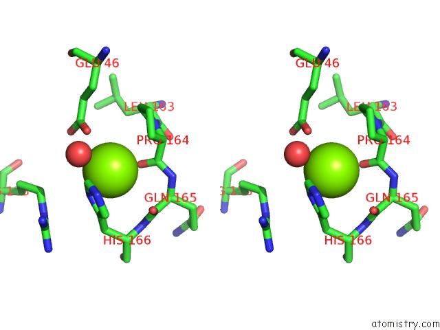
Magnesium binding site 1 out of 4 in 1jed
Go back to
Magnesium binding site 1 out
of 4 in the Crystal Structure of Atp Sulfurylase in Complex with Adp
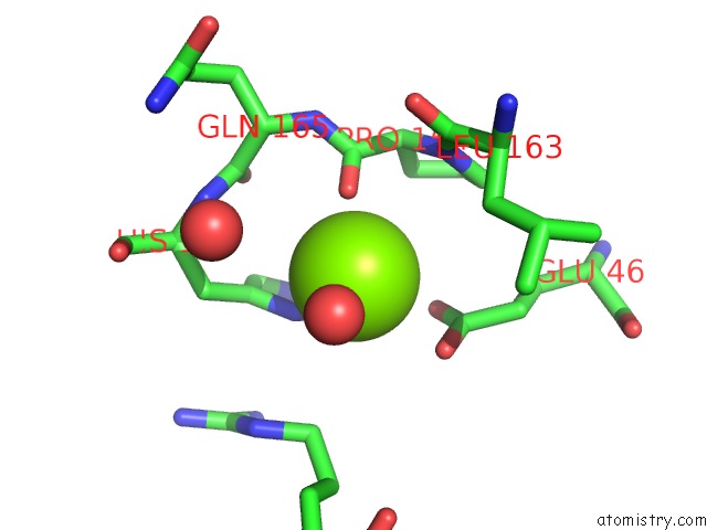
Mono view

Stereo pair view
Mono view
Stereo pair view
A full contact list of Magnesium with other atoms in the Mg binding
site number 1 of Crystal Structure of Atp Sulfurylase in Complex with Adp within 5.0Å range:
|
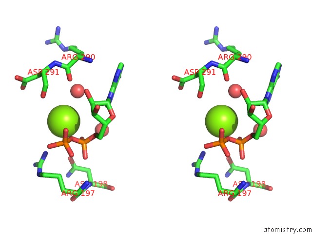
Magnesium binding site 2 out of 4 in 1jed
Go back to
Magnesium binding site 2 out
of 4 in the Crystal Structure of Atp Sulfurylase in Complex with Adp
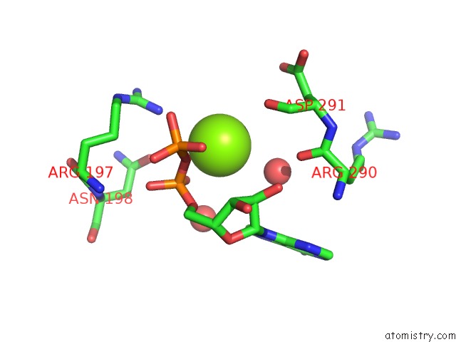
Mono view
Stereo pair view
Mono view
Stereo pair view
A full contact list of Magnesium with other atoms in the Mg binding
site number 2 of Crystal Structure of Atp Sulfurylase in Complex with Adp within 5.0Å range:
|
Magnesium binding site 3 out of 4 in 1jed
Go back to
Magnesium binding site 3 out
of 4 in the Crystal Structure of Atp Sulfurylase in Complex with Adp

Mono view
Stereo pair view
Mono view
Stereo pair view
A full contact list of Magnesium with other atoms in the Mg binding
site number 3 of Crystal Structure of Atp Sulfurylase in Complex with Adp within 5.0Å range:
|
Magnesium binding site 4 out of 4 in 1jed
Go back to
Magnesium binding site 4 out
of 4 in the Crystal Structure of Atp Sulfurylase in Complex with Adp

Mono view

Stereo pair view
Mono view
Stereo pair view
A full contact list of Magnesium with other atoms in the Mg binding
site number 4 of Crystal Structure of Atp Sulfurylase in Complex with Adp within 5.0Å range:
|
Reference:
T.C.Ullrich,
R.Huber.
The Complex Structures of Atp Sulfurylase with Thiosulfate, Adp and Chlorate Reveal New Insights in Inhibitory Effects and the Catalytic Cycle. J.Mol.Biol. V. 313 1117 2001.
ISSN: ISSN 0022-2836
PubMed: 11700067
DOI: 10.1006/JMBI.2001.5098
Page generated: Tue Aug 13 06:32:57 2024
ISSN: ISSN 0022-2836
PubMed: 11700067
DOI: 10.1006/JMBI.2001.5098
Last articles
Cl in 5WKKCl in 5WKI
Cl in 5WJO
Cl in 5WKE
Cl in 5WKG
Cl in 5WHV
Cl in 5WJL
Cl in 5WJK
Cl in 5WJI
Cl in 5WIV