Magnesium »
PDB 1jcg-1jwy »
1jls »
Magnesium in PDB 1jls: Structure of the Uracil Phosphoribosyltransferase Uracil/Cpr 2 Mutant C128V
Enzymatic activity of Structure of the Uracil Phosphoribosyltransferase Uracil/Cpr 2 Mutant C128V
All present enzymatic activity of Structure of the Uracil Phosphoribosyltransferase Uracil/Cpr 2 Mutant C128V:
2.4.2.9;
2.4.2.9;
Protein crystallography data
The structure of Structure of the Uracil Phosphoribosyltransferase Uracil/Cpr 2 Mutant C128V, PDB code: 1jls
was solved by
M.A.Schumacher,
C.J.Bashor,
K.Otsu,
S.Zu,
R.Parry,
B.Ullman,
R.G.Brennan,
with X-Ray Crystallography technique. A brief refinement statistics is given in the table below:
| Resolution Low / High (Å) | 60.03 / 2.50 |
| Space group | P 1 21 1 |
| Cell size a, b, c (Å), α, β, γ (°) | 71.400, 111.400, 71.900, 90.00, 97.70, 90.00 |
| R / Rfree (%) | 23.2 / 28.8 |
Magnesium Binding Sites:
The binding sites of Magnesium atom in the Structure of the Uracil Phosphoribosyltransferase Uracil/Cpr 2 Mutant C128V
(pdb code 1jls). This binding sites where shown within
5.0 Angstroms radius around Magnesium atom.
In total only one binding site of Magnesium was determined in the Structure of the Uracil Phosphoribosyltransferase Uracil/Cpr 2 Mutant C128V, PDB code: 1jls:
In total only one binding site of Magnesium was determined in the Structure of the Uracil Phosphoribosyltransferase Uracil/Cpr 2 Mutant C128V, PDB code: 1jls:
Magnesium binding site 1 out of 1 in 1jls
Go back to
Magnesium binding site 1 out
of 1 in the Structure of the Uracil Phosphoribosyltransferase Uracil/Cpr 2 Mutant C128V
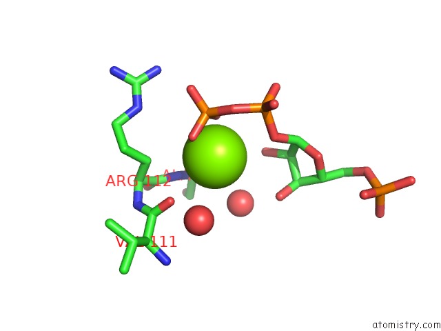
Mono view
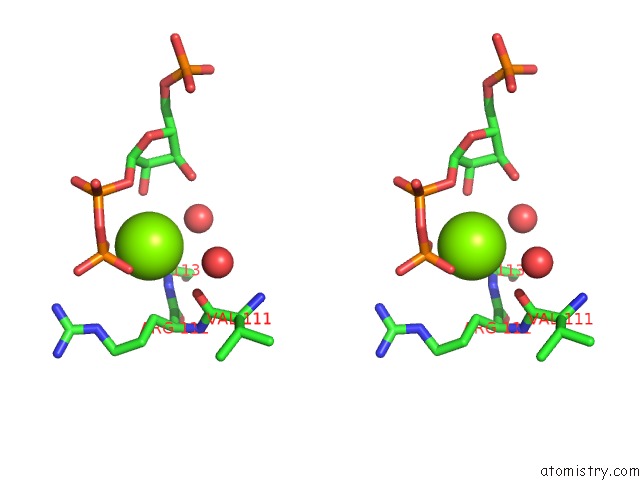
Stereo pair view
Mono view
Stereo pair view
A full contact list of Magnesium with other atoms in the Mg binding
site number 1 of Structure of the Uracil Phosphoribosyltransferase Uracil/Cpr 2 Mutant C128V within 5.0Å range:
|
Reference:
M.A.Schumacher,
C.J.Bashor,
M.H.Song,
K.Otsu,
S.Zhu,
R.J.Parry,
B.Ullman,
R.G.Brennan.
The Structural Mechanism of Gtp Stabilized Oligomerization and Catalytic Activation of the Toxoplasma Gondii Uracil Phosphoribosyltransferase. Proc.Natl.Acad.Sci.Usa V. 99 78 2002.
ISSN: ISSN 0027-8424
PubMed: 11773618
DOI: 10.1073/PNAS.012399599
Page generated: Sat Aug 9 23:23:07 2025
ISSN: ISSN 0027-8424
PubMed: 11773618
DOI: 10.1073/PNAS.012399599
Last articles
Mg in 1Q6TMg in 1Q6Z
Mg in 1Q78
Mg in 1Q6S
Mg in 1Q6R
Mg in 1Q6Q
Mg in 1Q6O
Mg in 1Q6N
Mg in 1Q3U
Mg in 1Q6L