Magnesium »
PDB 1jcg-1jwy »
1jm1 »
Magnesium in PDB 1jm1: Crystal Structure of the Soluble Domain of the Rieske Protein II (Soxf) From Sulfolobus Acidocaldarius
Protein crystallography data
The structure of Crystal Structure of the Soluble Domain of the Rieske Protein II (Soxf) From Sulfolobus Acidocaldarius, PDB code: 1jm1
was solved by
H.Boenisch,
C.L.Schmidt,
G.Schaefer,
R.Ladenstein,
with X-Ray Crystallography technique. A brief refinement statistics is given in the table below:
| Resolution Low / High (Å) | 20.00 / 1.11 |
| Space group | P 61 |
| Cell size a, b, c (Å), α, β, γ (°) | 80.252, 80.252, 75.624, 90.00, 90.00, 120.00 |
| R / Rfree (%) | 10.6 / 12.5 |
Other elements in 1jm1:
The structure of Crystal Structure of the Soluble Domain of the Rieske Protein II (Soxf) From Sulfolobus Acidocaldarius also contains other interesting chemical elements:
| Iron | (Fe) | 2 atoms |
Magnesium Binding Sites:
The binding sites of Magnesium atom in the Crystal Structure of the Soluble Domain of the Rieske Protein II (Soxf) From Sulfolobus Acidocaldarius
(pdb code 1jm1). This binding sites where shown within
5.0 Angstroms radius around Magnesium atom.
In total only one binding site of Magnesium was determined in the Crystal Structure of the Soluble Domain of the Rieske Protein II (Soxf) From Sulfolobus Acidocaldarius, PDB code: 1jm1:
In total only one binding site of Magnesium was determined in the Crystal Structure of the Soluble Domain of the Rieske Protein II (Soxf) From Sulfolobus Acidocaldarius, PDB code: 1jm1:
Magnesium binding site 1 out of 1 in 1jm1
Go back to
Magnesium binding site 1 out
of 1 in the Crystal Structure of the Soluble Domain of the Rieske Protein II (Soxf) From Sulfolobus Acidocaldarius
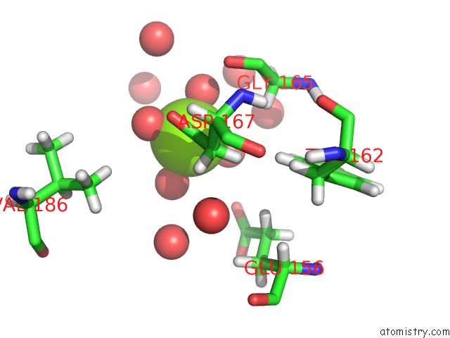
Mono view
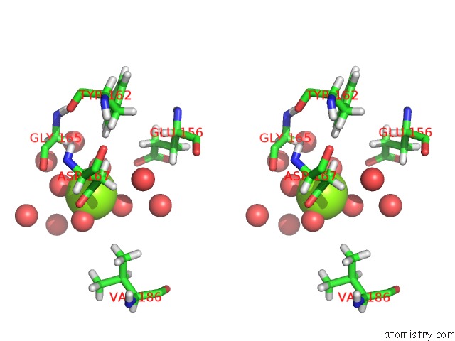
Stereo pair view
Mono view
Stereo pair view
A full contact list of Magnesium with other atoms in the Mg binding
site number 1 of Crystal Structure of the Soluble Domain of the Rieske Protein II (Soxf) From Sulfolobus Acidocaldarius within 5.0Å range:
|
Reference:
H.Bonisch,
C.L.Schmidt,
G.Schafer,
R.Ladenstein.
The Structure of the Soluble Domain of An Archaeal Rieske Iron-Sulfur Protein at 1.1 A Resolution. J.Mol.Biol. V. 319 791 2002.
ISSN: ISSN 0022-2836
PubMed: 12054871
DOI: 10.1016/S0022-2836(02)00323-6
Page generated: Tue Aug 13 06:37:21 2024
ISSN: ISSN 0022-2836
PubMed: 12054871
DOI: 10.1016/S0022-2836(02)00323-6
Last articles
Zn in 9J0NZn in 9J0O
Zn in 9J0P
Zn in 9FJX
Zn in 9EKB
Zn in 9C0F
Zn in 9CAH
Zn in 9CH0
Zn in 9CH3
Zn in 9CH1