Magnesium »
PDB 1jcg-1jwy »
1jms »
Magnesium in PDB 1jms: Crystal Structure of the Catalytic Core of Murine Terminal Deoxynucleotidyl Transferase
Enzymatic activity of Crystal Structure of the Catalytic Core of Murine Terminal Deoxynucleotidyl Transferase
All present enzymatic activity of Crystal Structure of the Catalytic Core of Murine Terminal Deoxynucleotidyl Transferase:
2.7.7.31;
2.7.7.31;
Protein crystallography data
The structure of Crystal Structure of the Catalytic Core of Murine Terminal Deoxynucleotidyl Transferase, PDB code: 1jms
was solved by
M.Delarue,
J.B.Boule,
J.Lescar,
N.Expert-Bezancon,
N.Sukumar,
N.Jourdan,
F.Rougeon,
C.Papanicolaou,
with X-Ray Crystallography technique. A brief refinement statistics is given in the table below:
| Resolution Low / High (Å) | 18.00 / 2.36 |
| Space group | P 21 21 21 |
| Cell size a, b, c (Å), α, β, γ (°) | 47.100, 85.200, 111.700, 90.00, 90.00, 90.00 |
| R / Rfree (%) | 21.4 / 25.9 |
Other elements in 1jms:
The structure of Crystal Structure of the Catalytic Core of Murine Terminal Deoxynucleotidyl Transferase also contains other interesting chemical elements:
| Sodium | (Na) | 1 atom |
Magnesium Binding Sites:
The binding sites of Magnesium atom in the Crystal Structure of the Catalytic Core of Murine Terminal Deoxynucleotidyl Transferase
(pdb code 1jms). This binding sites where shown within
5.0 Angstroms radius around Magnesium atom.
In total only one binding site of Magnesium was determined in the Crystal Structure of the Catalytic Core of Murine Terminal Deoxynucleotidyl Transferase, PDB code: 1jms:
In total only one binding site of Magnesium was determined in the Crystal Structure of the Catalytic Core of Murine Terminal Deoxynucleotidyl Transferase, PDB code: 1jms:
Magnesium binding site 1 out of 1 in 1jms
Go back to
Magnesium binding site 1 out
of 1 in the Crystal Structure of the Catalytic Core of Murine Terminal Deoxynucleotidyl Transferase
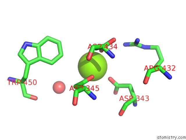
Mono view
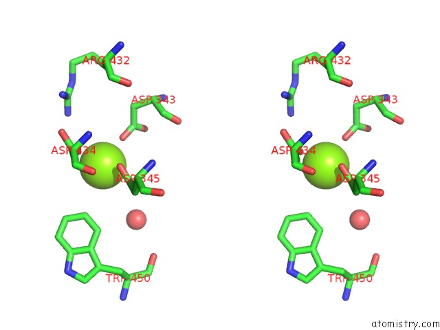
Stereo pair view
Mono view
Stereo pair view
A full contact list of Magnesium with other atoms in the Mg binding
site number 1 of Crystal Structure of the Catalytic Core of Murine Terminal Deoxynucleotidyl Transferase within 5.0Å range:
|
Reference:
M.Delarue,
J.B.Boule,
J.Lescar,
N.Expert-Bezancon,
N.Jourdan,
N.Sukumar,
F.Rougeon,
C.Papanicolaou.
Crystal Structures of A Template-Independent Dna Polymerase: Murine Terminal Deoxynucleotidyltransferase. Embo J. V. 21 427 2002.
ISSN: ISSN 0261-4189
PubMed: 11823435
DOI: 10.1093/EMBOJ/21.3.427
Page generated: Tue Aug 13 06:37:33 2024
ISSN: ISSN 0261-4189
PubMed: 11823435
DOI: 10.1093/EMBOJ/21.3.427
Last articles
Zn in 9J0NZn in 9J0O
Zn in 9J0P
Zn in 9FJX
Zn in 9EKB
Zn in 9C0F
Zn in 9CAH
Zn in 9CH0
Zn in 9CH3
Zn in 9CH1