Magnesium »
PDB 1jx2-1k9m »
1jx2 »
Magnesium in PDB 1jx2: Crystal Structure of the Nucleotide-Free Dynamin A Gtpase Domain, Determined As Myosin Fusion
Protein crystallography data
The structure of Crystal Structure of the Nucleotide-Free Dynamin A Gtpase Domain, Determined As Myosin Fusion, PDB code: 1jx2
was solved by
H.H.Niemann,
M.L.W.Knetsch,
A.Scherer,
D.J.Manstein,
F.J.Kull,
with X-Ray Crystallography technique. A brief refinement statistics is given in the table below:
| Resolution Low / High (Å) | 14.96 / 2.30 |
| Space group | P 1 21 1 |
| Cell size a, b, c (Å), α, β, γ (°) | 54.450, 62.040, 181.200, 90.00, 94.79, 90.00 |
| R / Rfree (%) | 19.7 / 25.5 |
Magnesium Binding Sites:
The binding sites of Magnesium atom in the Crystal Structure of the Nucleotide-Free Dynamin A Gtpase Domain, Determined As Myosin Fusion
(pdb code 1jx2). This binding sites where shown within
5.0 Angstroms radius around Magnesium atom.
In total only one binding site of Magnesium was determined in the Crystal Structure of the Nucleotide-Free Dynamin A Gtpase Domain, Determined As Myosin Fusion, PDB code: 1jx2:
In total only one binding site of Magnesium was determined in the Crystal Structure of the Nucleotide-Free Dynamin A Gtpase Domain, Determined As Myosin Fusion, PDB code: 1jx2:
Magnesium binding site 1 out of 1 in 1jx2
Go back to
Magnesium binding site 1 out
of 1 in the Crystal Structure of the Nucleotide-Free Dynamin A Gtpase Domain, Determined As Myosin Fusion
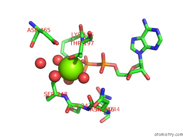
Mono view
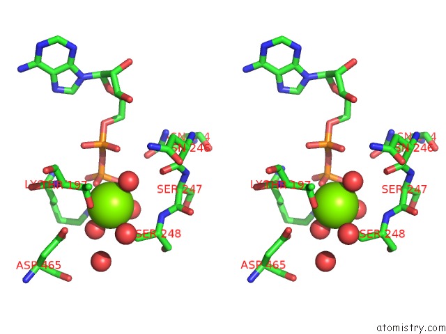
Stereo pair view
Mono view
Stereo pair view
A full contact list of Magnesium with other atoms in the Mg binding
site number 1 of Crystal Structure of the Nucleotide-Free Dynamin A Gtpase Domain, Determined As Myosin Fusion within 5.0Å range:
|
Reference:
H.H.Niemann,
M.L.Knetsch,
A.Scherer,
D.J.Manstein,
F.J.Kull.
Crystal Structure of A Dynamin Gtpase Domain in Both Nucleotide-Free and Gdp-Bound Forms. Embo J. V. 20 5813 2001.
ISSN: ISSN 0261-4189
PubMed: 11689422
DOI: 10.1093/EMBOJ/20.21.5813
Page generated: Sat Aug 9 23:32:24 2025
ISSN: ISSN 0261-4189
PubMed: 11689422
DOI: 10.1093/EMBOJ/20.21.5813
Last articles
Mg in 3I5XMg in 3I4K
Mg in 3I5F
Mg in 3I5C
Mg in 3I4N
Mg in 3I3E
Mg in 3I4D
Mg in 3I4M
Mg in 3I3D
Mg in 3I3B