Magnesium »
PDB 1jx2-1k9m »
1k4i »
Magnesium in PDB 1k4i: Crystal Structure of 3,4-Dihydroxy-2-Butanone 4-Phosphate Synthase in Complex with Two Magnesium Ions
Protein crystallography data
The structure of Crystal Structure of 3,4-Dihydroxy-2-Butanone 4-Phosphate Synthase in Complex with Two Magnesium Ions, PDB code: 1k4i
was solved by
D.-I.Liao,
Y.-J.Zheng,
P.V.Viitanen,
D.B.Jordan,
with X-Ray Crystallography technique. A brief refinement statistics is given in the table below:
| Resolution Low / High (Å) | 25.00 / 0.98 |
| Space group | P 21 21 2 |
| Cell size a, b, c (Å), α, β, γ (°) | 53.889, 88.147, 43.995, 90.00, 90.00, 90.00 |
| R / Rfree (%) | n/a / n/a |
Magnesium Binding Sites:
The binding sites of Magnesium atom in the Crystal Structure of 3,4-Dihydroxy-2-Butanone 4-Phosphate Synthase in Complex with Two Magnesium Ions
(pdb code 1k4i). This binding sites where shown within
5.0 Angstroms radius around Magnesium atom.
In total 2 binding sites of Magnesium where determined in the Crystal Structure of 3,4-Dihydroxy-2-Butanone 4-Phosphate Synthase in Complex with Two Magnesium Ions, PDB code: 1k4i:
Jump to Magnesium binding site number: 1; 2;
In total 2 binding sites of Magnesium where determined in the Crystal Structure of 3,4-Dihydroxy-2-Butanone 4-Phosphate Synthase in Complex with Two Magnesium Ions, PDB code: 1k4i:
Jump to Magnesium binding site number: 1; 2;
Magnesium binding site 1 out of 2 in 1k4i
Go back to
Magnesium binding site 1 out
of 2 in the Crystal Structure of 3,4-Dihydroxy-2-Butanone 4-Phosphate Synthase in Complex with Two Magnesium Ions
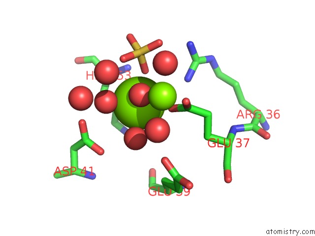
Mono view
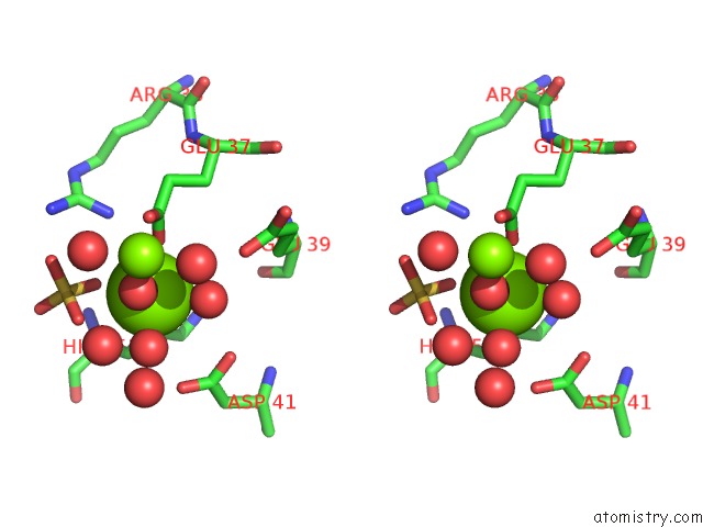
Stereo pair view
Mono view
Stereo pair view
A full contact list of Magnesium with other atoms in the Mg binding
site number 1 of Crystal Structure of 3,4-Dihydroxy-2-Butanone 4-Phosphate Synthase in Complex with Two Magnesium Ions within 5.0Å range:
|
Magnesium binding site 2 out of 2 in 1k4i
Go back to
Magnesium binding site 2 out
of 2 in the Crystal Structure of 3,4-Dihydroxy-2-Butanone 4-Phosphate Synthase in Complex with Two Magnesium Ions
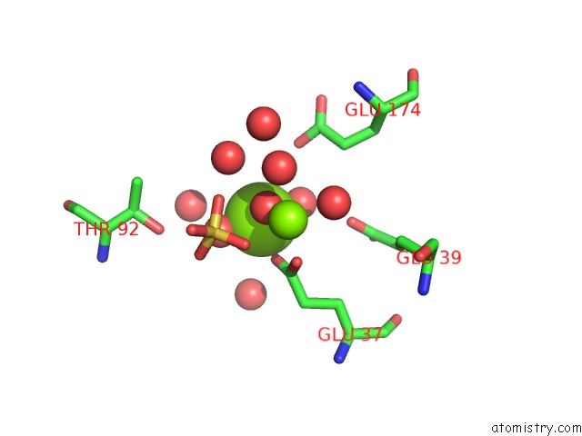
Mono view
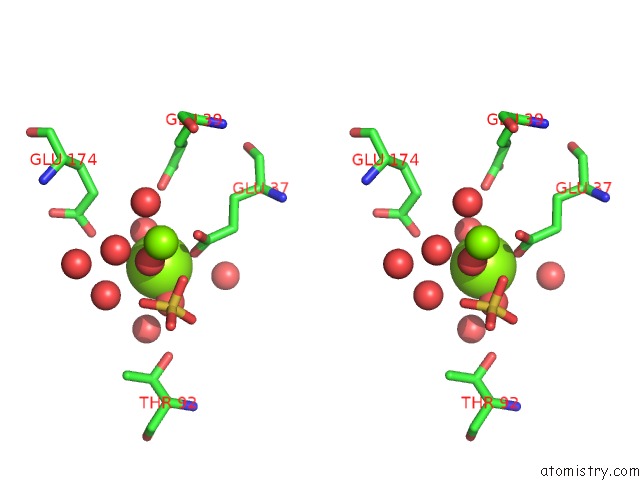
Stereo pair view
Mono view
Stereo pair view
A full contact list of Magnesium with other atoms in the Mg binding
site number 2 of Crystal Structure of 3,4-Dihydroxy-2-Butanone 4-Phosphate Synthase in Complex with Two Magnesium Ions within 5.0Å range:
|
Reference:
D.I.Liao,
Y.J.Zheng,
P.V.Viitanen,
D.B.Jordan.
Structural Definition of the Active Site and Catalytic Mechanism of 3,4-Dihydroxy-2-Butanone-4-Phosphate Synthase. Biochemistry V. 41 1795 2002.
ISSN: ISSN 0006-2960
PubMed: 11827524
DOI: 10.1021/BI015652U
Page generated: Tue Aug 13 07:06:45 2024
ISSN: ISSN 0006-2960
PubMed: 11827524
DOI: 10.1021/BI015652U
Last articles
Fe in 2YXOFe in 2YRS
Fe in 2YXC
Fe in 2YNM
Fe in 2YVJ
Fe in 2YP1
Fe in 2YU2
Fe in 2YU1
Fe in 2YQB
Fe in 2YOO