Magnesium »
PDB 1jx2-1k9m »
1k6d »
Magnesium in PDB 1k6d: Crystal Structure of Acetate Coa-Transferase Alpha Subunit
Enzymatic activity of Crystal Structure of Acetate Coa-Transferase Alpha Subunit
All present enzymatic activity of Crystal Structure of Acetate Coa-Transferase Alpha Subunit:
2.8.3.8;
2.8.3.8;
Protein crystallography data
The structure of Crystal Structure of Acetate Coa-Transferase Alpha Subunit, PDB code: 1k6d
was solved by
S.Korolev,
O.Koroleva,
K.Petterson,
F.Collart,
I.Dementieva,
A.Joachimiak,
Midwest Center For Structural Genomics (Mcsg),
with X-Ray Crystallography technique. A brief refinement statistics is given in the table below:
| Resolution Low / High (Å) | 36.00 / 1.90 |
| Space group | P 62 |
| Cell size a, b, c (Å), α, β, γ (°) | 88.496, 88.496, 105.080, 90.00, 90.00, 120.00 |
| R / Rfree (%) | 20.1 / 23.5 |
Magnesium Binding Sites:
The binding sites of Magnesium atom in the Crystal Structure of Acetate Coa-Transferase Alpha Subunit
(pdb code 1k6d). This binding sites where shown within
5.0 Angstroms radius around Magnesium atom.
In total 4 binding sites of Magnesium where determined in the Crystal Structure of Acetate Coa-Transferase Alpha Subunit, PDB code: 1k6d:
Jump to Magnesium binding site number: 1; 2; 3; 4;
In total 4 binding sites of Magnesium where determined in the Crystal Structure of Acetate Coa-Transferase Alpha Subunit, PDB code: 1k6d:
Jump to Magnesium binding site number: 1; 2; 3; 4;
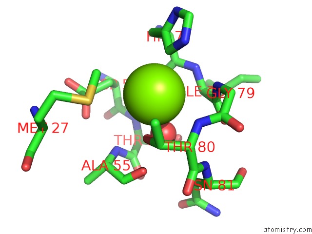
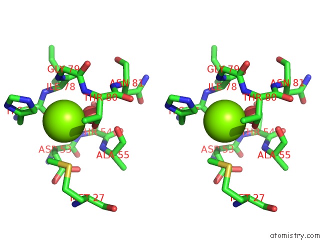
Magnesium binding site 1 out of 4 in 1k6d
Go back to
Magnesium binding site 1 out
of 4 in the Crystal Structure of Acetate Coa-Transferase Alpha Subunit
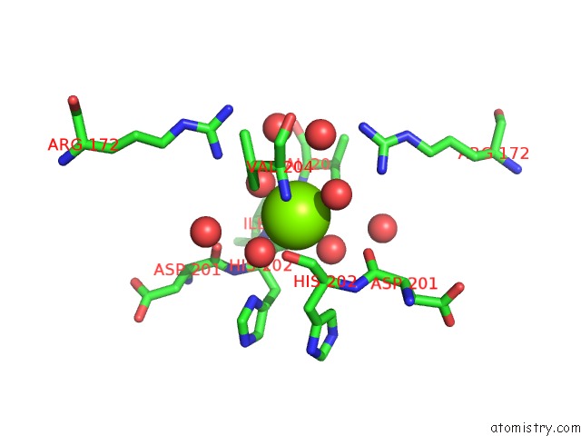
Mono view
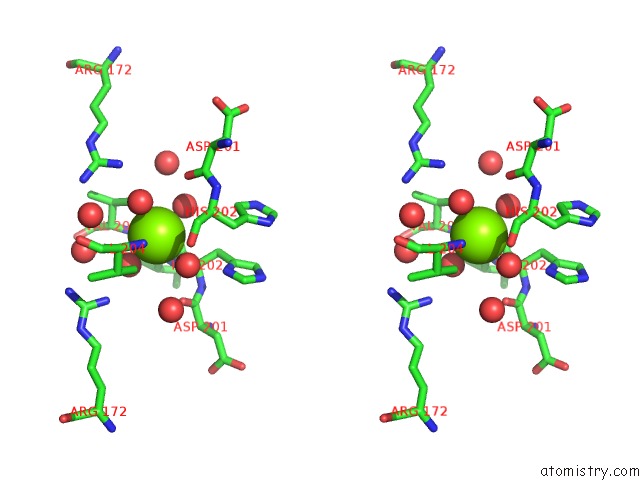
Stereo pair view
Mono view
Stereo pair view
A full contact list of Magnesium with other atoms in the Mg binding
site number 1 of Crystal Structure of Acetate Coa-Transferase Alpha Subunit within 5.0Å range:
|
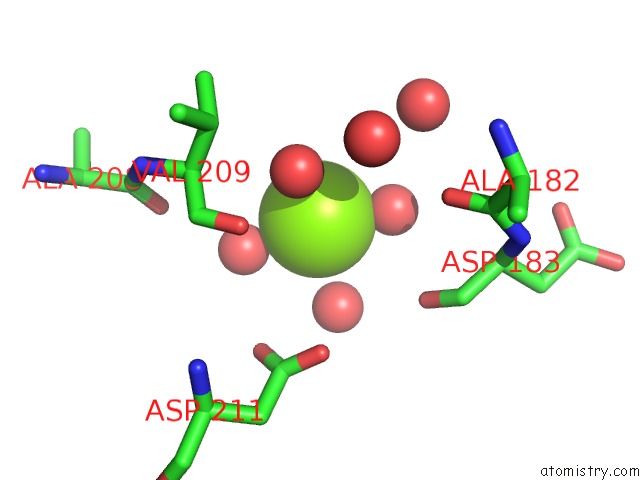
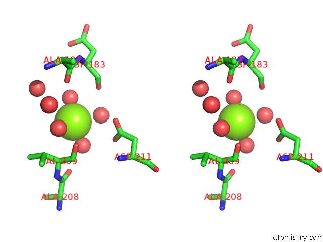
Magnesium binding site 2 out of 4 in 1k6d
Go back to
Magnesium binding site 2 out
of 4 in the Crystal Structure of Acetate Coa-Transferase Alpha Subunit
Mono view
Stereo pair view
Mono view
Stereo pair view
A full contact list of Magnesium with other atoms in the Mg binding
site number 2 of Crystal Structure of Acetate Coa-Transferase Alpha Subunit within 5.0Å range:
|
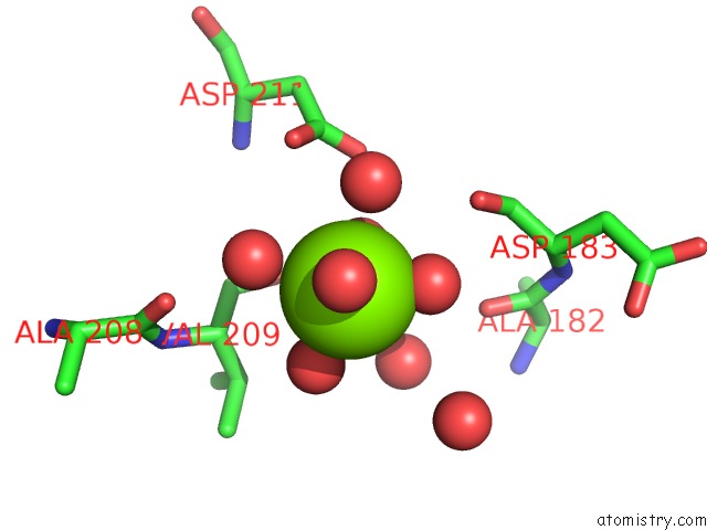
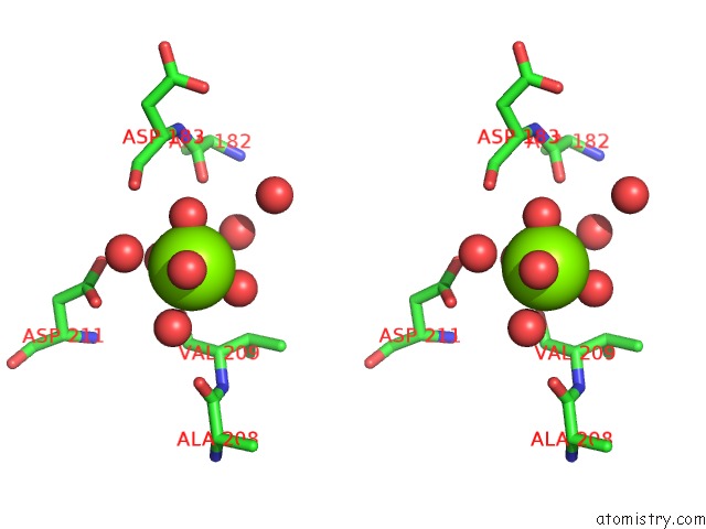
Magnesium binding site 3 out of 4 in 1k6d
Go back to
Magnesium binding site 3 out
of 4 in the Crystal Structure of Acetate Coa-Transferase Alpha Subunit
Mono view
Stereo pair view
Mono view
Stereo pair view
A full contact list of Magnesium with other atoms in the Mg binding
site number 3 of Crystal Structure of Acetate Coa-Transferase Alpha Subunit within 5.0Å range:
|
Magnesium binding site 4 out of 4 in 1k6d
Go back to
Magnesium binding site 4 out
of 4 in the Crystal Structure of Acetate Coa-Transferase Alpha Subunit
Mono view
Stereo pair view
Mono view
Stereo pair view
A full contact list of Magnesium with other atoms in the Mg binding
site number 4 of Crystal Structure of Acetate Coa-Transferase Alpha Subunit within 5.0Å range:
|
Reference:
S.Korolev,
O.Koroleva,
K.Petterson,
M.Gu,
F.Collart,
I.Dementieva,
A.Joachimiak.
Autotracing of Escherichia Coli Acetate Coa-Transferase Alpha-Subunit Structure Using 3.4 A Mad and 1.9 A Native Data. Acta Crystallogr.,Sect.D V. 58 2116 2002.
ISSN: ISSN 0907-4449
PubMed: 12454473
DOI: 10.1107/S0907444902017055
Page generated: Tue Aug 13 07:08:26 2024
ISSN: ISSN 0907-4449
PubMed: 12454473
DOI: 10.1107/S0907444902017055
Last articles
Zn in 9MJ5Zn in 9HNW
Zn in 9G0L
Zn in 9FNE
Zn in 9DZN
Zn in 9E0I
Zn in 9D32
Zn in 9DAK
Zn in 8ZXC
Zn in 8ZUF