Magnesium »
PDB 1jx2-1k9m »
1k77 »
Magnesium in PDB 1k77: Crystal Structure of EC1530, A Putative Oxygenase From Escherichia Coli
Protein crystallography data
The structure of Crystal Structure of EC1530, A Putative Oxygenase From Escherichia Coli, PDB code: 1k77
was solved by
Y.Kim,
T.Skarina,
S.Beasley,
R.Laskowski,
C.H.Arrowsmith,
A.Joachimiak,
A.M.Edwards,
A.Savchenko,
Midwest Center For Structural Genomics(Mcsg),
with X-Ray Crystallography technique. A brief refinement statistics is given in the table below:
| Resolution Low / High (Å) | 38.91 / 1.63 |
| Space group | C 1 2 1 |
| Cell size a, b, c (Å), α, β, γ (°) | 104.907, 74.368, 39.376, 90.00, 98.81, 90.00 |
| R / Rfree (%) | 19.4 / 21.4 |
Magnesium Binding Sites:
The binding sites of Magnesium atom in the Crystal Structure of EC1530, A Putative Oxygenase From Escherichia Coli
(pdb code 1k77). This binding sites where shown within
5.0 Angstroms radius around Magnesium atom.
In total 2 binding sites of Magnesium where determined in the Crystal Structure of EC1530, A Putative Oxygenase From Escherichia Coli, PDB code: 1k77:
Jump to Magnesium binding site number: 1; 2;
In total 2 binding sites of Magnesium where determined in the Crystal Structure of EC1530, A Putative Oxygenase From Escherichia Coli, PDB code: 1k77:
Jump to Magnesium binding site number: 1; 2;
Magnesium binding site 1 out of 2 in 1k77
Go back to
Magnesium binding site 1 out
of 2 in the Crystal Structure of EC1530, A Putative Oxygenase From Escherichia Coli
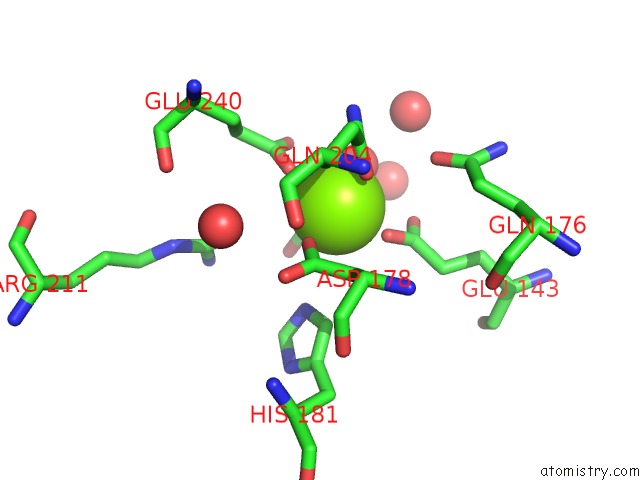
Mono view
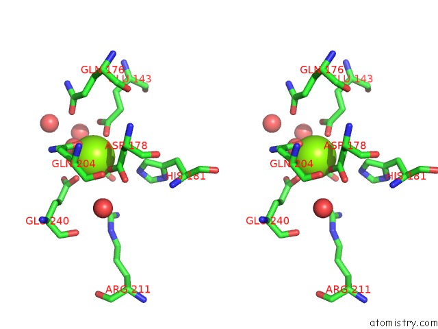
Stereo pair view
Mono view
Stereo pair view
A full contact list of Magnesium with other atoms in the Mg binding
site number 1 of Crystal Structure of EC1530, A Putative Oxygenase From Escherichia Coli within 5.0Å range:
|
Magnesium binding site 2 out of 2 in 1k77
Go back to
Magnesium binding site 2 out
of 2 in the Crystal Structure of EC1530, A Putative Oxygenase From Escherichia Coli
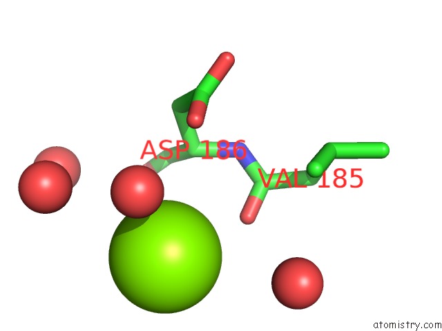
Mono view
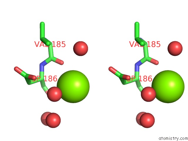
Stereo pair view
Mono view
Stereo pair view
A full contact list of Magnesium with other atoms in the Mg binding
site number 2 of Crystal Structure of EC1530, A Putative Oxygenase From Escherichia Coli within 5.0Å range:
|
Reference:
Y.Kim,
T.Skarina,
S.Beasley,
R.Laskowski,
C.H.Arrowsmith,
A.Joachimiak,
A.M.Edwards,
A.Savchenko.
Crystal Structure of Escherichia Coli EC1530, A Glyoxylate Induced Protein Ygbm. Proteins V. 48 427 2002.
ISSN: ISSN 0887-3585
PubMed: 12112708
DOI: 10.1002/PROT.10160
Page generated: Tue Aug 13 07:09:53 2024
ISSN: ISSN 0887-3585
PubMed: 12112708
DOI: 10.1002/PROT.10160
Last articles
Fe in 2YXOFe in 2YRS
Fe in 2YXC
Fe in 2YNM
Fe in 2YVJ
Fe in 2YP1
Fe in 2YU2
Fe in 2YU1
Fe in 2YQB
Fe in 2YOO