Magnesium »
PDB 1k9w-1kk3 »
1kdn »
Magnesium in PDB 1kdn: Structure of Nucleoside Diphosphate Kinase
Enzymatic activity of Structure of Nucleoside Diphosphate Kinase
All present enzymatic activity of Structure of Nucleoside Diphosphate Kinase:
2.7.4.6;
2.7.4.6;
Protein crystallography data
The structure of Structure of Nucleoside Diphosphate Kinase, PDB code: 1kdn
was solved by
J.Cherfils,
Y.W.Xu,
S.Morera,
J.Janin,
with X-Ray Crystallography technique. A brief refinement statistics is given in the table below:
| Resolution Low / High (Å) | N/A / 2.00 |
| Space group | P 31 2 1 |
| Cell size a, b, c (Å), α, β, γ (°) | 71.354, 71.354, 153.494, 90.00, 90.00, 120.00 |
| R / Rfree (%) | 17.7 / 17.9 |
Other elements in 1kdn:
The structure of Structure of Nucleoside Diphosphate Kinase also contains other interesting chemical elements:
| Fluorine | (F) | 9 atoms |
| Aluminium | (Al) | 3 atoms |
Magnesium Binding Sites:
The binding sites of Magnesium atom in the Structure of Nucleoside Diphosphate Kinase
(pdb code 1kdn). This binding sites where shown within
5.0 Angstroms radius around Magnesium atom.
In total 3 binding sites of Magnesium where determined in the Structure of Nucleoside Diphosphate Kinase, PDB code: 1kdn:
Jump to Magnesium binding site number: 1; 2; 3;
In total 3 binding sites of Magnesium where determined in the Structure of Nucleoside Diphosphate Kinase, PDB code: 1kdn:
Jump to Magnesium binding site number: 1; 2; 3;
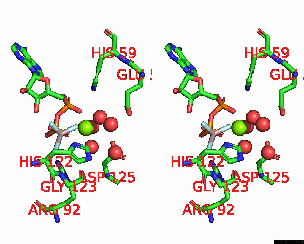
Magnesium binding site 1 out of 3 in 1kdn
Go back to
Magnesium binding site 1 out
of 3 in the Structure of Nucleoside Diphosphate Kinase
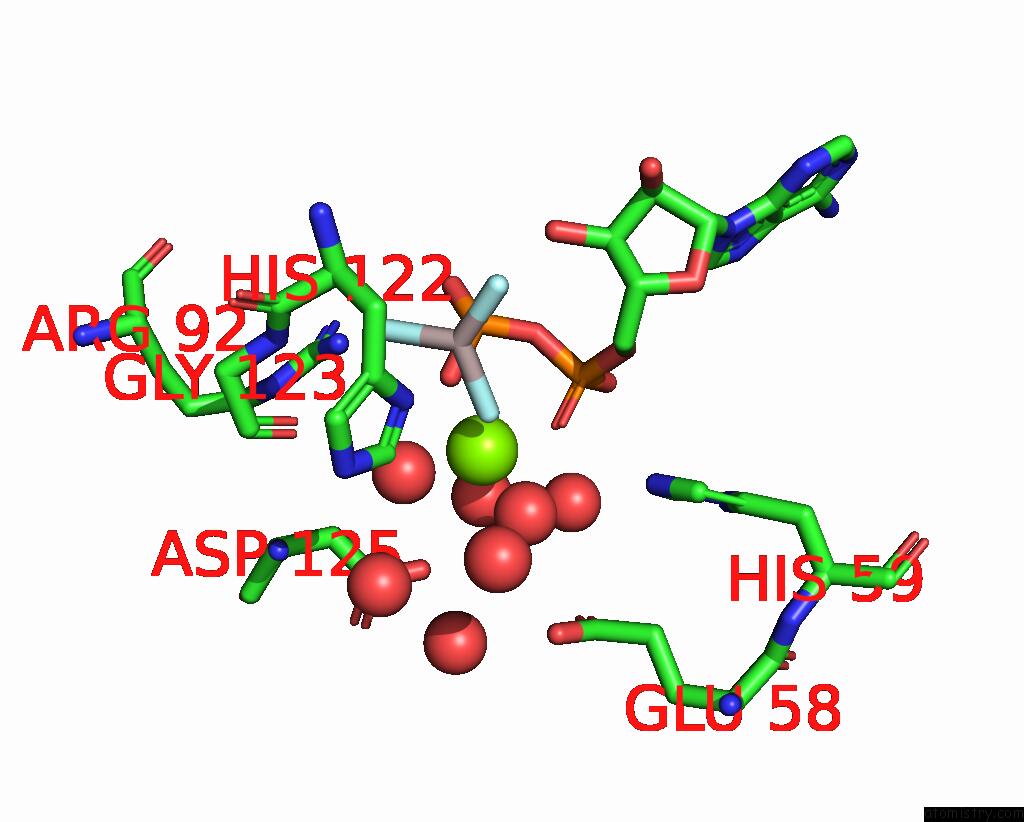
Mono view
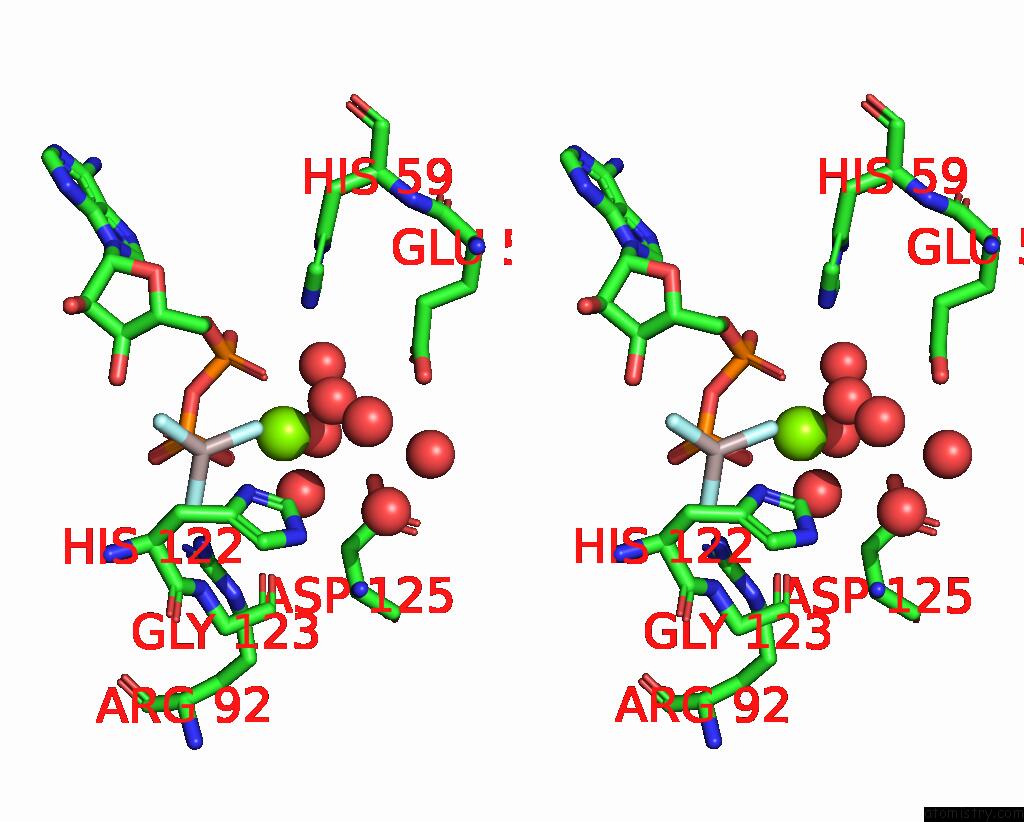
Stereo pair view
Mono view
Stereo pair view
A full contact list of Magnesium with other atoms in the Mg binding
site number 1 of Structure of Nucleoside Diphosphate Kinase within 5.0Å range:
|
Magnesium binding site 2 out of 3 in 1kdn
Go back to
Magnesium binding site 2 out
of 3 in the Structure of Nucleoside Diphosphate Kinase
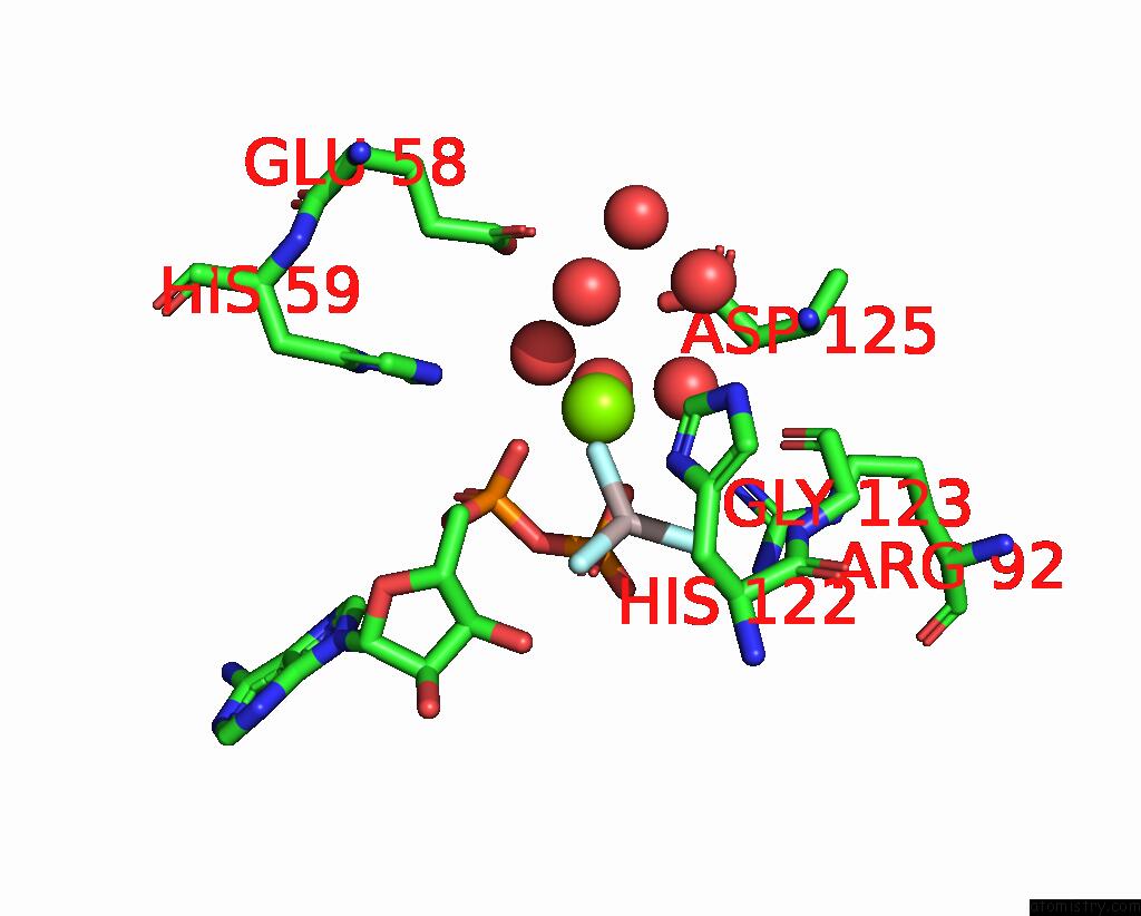
Mono view
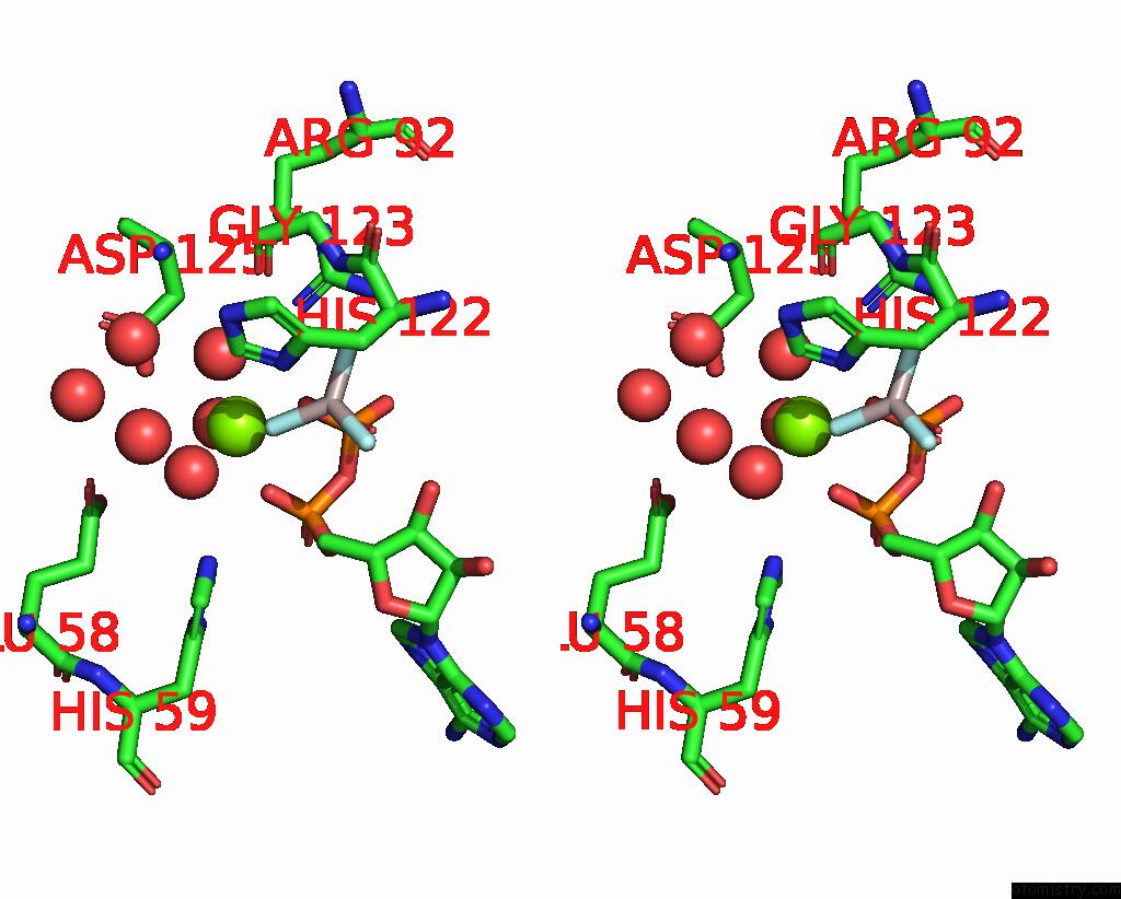
Stereo pair view
Mono view
Stereo pair view
A full contact list of Magnesium with other atoms in the Mg binding
site number 2 of Structure of Nucleoside Diphosphate Kinase within 5.0Å range:
|
Magnesium binding site 3 out of 3 in 1kdn
Go back to
Magnesium binding site 3 out
of 3 in the Structure of Nucleoside Diphosphate Kinase

Mono view
Stereo pair view
Mono view
Stereo pair view
A full contact list of Magnesium with other atoms in the Mg binding
site number 3 of Structure of Nucleoside Diphosphate Kinase within 5.0Å range:
|
Reference:
Y.W.Xu,
S.Morera,
J.Janin,
J.Cherfils.
ALF3 Mimics the Transition State of Protein Phosphorylation in the Crystal Structure of Nucleoside Diphosphate Kinase and Mgadp. Proc.Natl.Acad.Sci.Usa V. 94 3579 1997.
ISSN: ISSN 0027-8424
PubMed: 9108019
DOI: 10.1073/PNAS.94.8.3579
Page generated: Tue Aug 13 07:41:31 2024
ISSN: ISSN 0027-8424
PubMed: 9108019
DOI: 10.1073/PNAS.94.8.3579
Last articles
Zn in 9J0NZn in 9J0O
Zn in 9J0P
Zn in 9FJX
Zn in 9EKB
Zn in 9C0F
Zn in 9CAH
Zn in 9CH0
Zn in 9CH3
Zn in 9CH1