Magnesium »
PDB 1k9w-1kk3 »
1kfg »
Magnesium in PDB 1kfg: The X-Ray Crystal Structure of CEL9G From Clostridium Cellulolyticum Complexed with A Thio-Oligosaccharide Inhibitor
Enzymatic activity of The X-Ray Crystal Structure of CEL9G From Clostridium Cellulolyticum Complexed with A Thio-Oligosaccharide Inhibitor
All present enzymatic activity of The X-Ray Crystal Structure of CEL9G From Clostridium Cellulolyticum Complexed with A Thio-Oligosaccharide Inhibitor:
3.2.1.4;
3.2.1.4;
Protein crystallography data
The structure of The X-Ray Crystal Structure of CEL9G From Clostridium Cellulolyticum Complexed with A Thio-Oligosaccharide Inhibitor, PDB code: 1kfg
was solved by
D.Mandelman,
A.Belaich,
J.-P.Belaich,
H.Driguez,
R.Haser,
with X-Ray Crystallography technique. A brief refinement statistics is given in the table below:
| Resolution Low / High (Å) | 42.42 / 1.90 |
| Space group | P 1 |
| Cell size a, b, c (Å), α, β, γ (°) | 57.040, 57.730, 86.630, 93.86, 100.83, 99.54 |
| R / Rfree (%) | 16.7 / 20.4 |
Other elements in 1kfg:
The structure of The X-Ray Crystal Structure of CEL9G From Clostridium Cellulolyticum Complexed with A Thio-Oligosaccharide Inhibitor also contains other interesting chemical elements:
| Nickel | (Ni) | 1 atom |
| Calcium | (Ca) | 4 atoms |
Magnesium Binding Sites:
The binding sites of Magnesium atom in the The X-Ray Crystal Structure of CEL9G From Clostridium Cellulolyticum Complexed with A Thio-Oligosaccharide Inhibitor
(pdb code 1kfg). This binding sites where shown within
5.0 Angstroms radius around Magnesium atom.
In total 5 binding sites of Magnesium where determined in the The X-Ray Crystal Structure of CEL9G From Clostridium Cellulolyticum Complexed with A Thio-Oligosaccharide Inhibitor, PDB code: 1kfg:
Jump to Magnesium binding site number: 1; 2; 3; 4; 5;
In total 5 binding sites of Magnesium where determined in the The X-Ray Crystal Structure of CEL9G From Clostridium Cellulolyticum Complexed with A Thio-Oligosaccharide Inhibitor, PDB code: 1kfg:
Jump to Magnesium binding site number: 1; 2; 3; 4; 5;
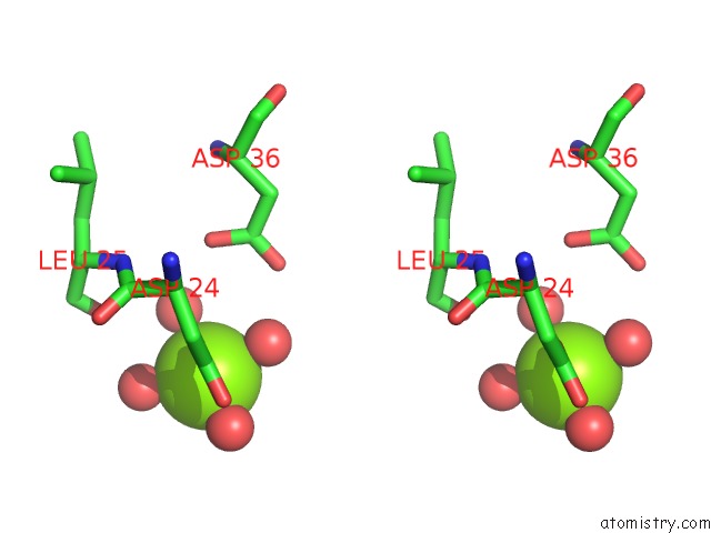
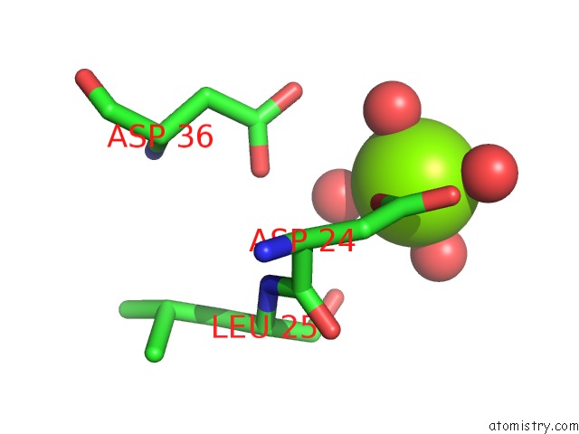
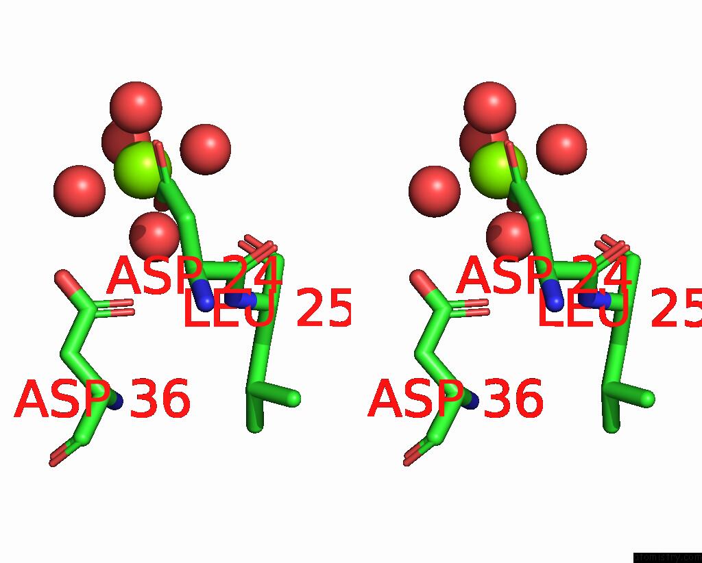
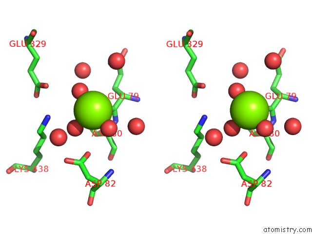
Magnesium binding site 1 out of 5 in 1kfg
Go back to
Magnesium binding site 1 out
of 5 in the The X-Ray Crystal Structure of CEL9G From Clostridium Cellulolyticum Complexed with A Thio-Oligosaccharide Inhibitor
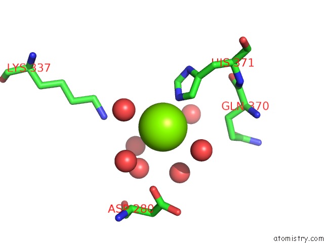
Mono view
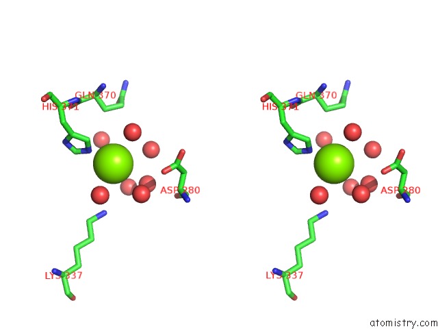
Stereo pair view
Mono view
Stereo pair view
A full contact list of Magnesium with other atoms in the Mg binding
site number 1 of The X-Ray Crystal Structure of CEL9G From Clostridium Cellulolyticum Complexed with A Thio-Oligosaccharide Inhibitor within 5.0Å range:
|
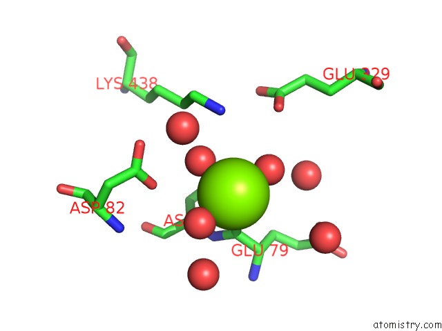
Magnesium binding site 2 out of 5 in 1kfg
Go back to
Magnesium binding site 2 out
of 5 in the The X-Ray Crystal Structure of CEL9G From Clostridium Cellulolyticum Complexed with A Thio-Oligosaccharide Inhibitor

Mono view
Stereo pair view
Mono view
Stereo pair view
A full contact list of Magnesium with other atoms in the Mg binding
site number 2 of The X-Ray Crystal Structure of CEL9G From Clostridium Cellulolyticum Complexed with A Thio-Oligosaccharide Inhibitor within 5.0Å range:
|
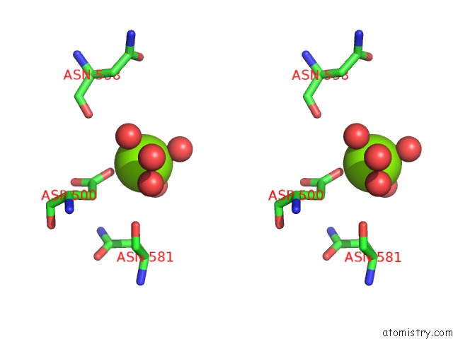
Magnesium binding site 3 out of 5 in 1kfg
Go back to
Magnesium binding site 3 out
of 5 in the The X-Ray Crystal Structure of CEL9G From Clostridium Cellulolyticum Complexed with A Thio-Oligosaccharide Inhibitor
Mono view
Stereo pair view
Mono view
Stereo pair view
A full contact list of Magnesium with other atoms in the Mg binding
site number 3 of The X-Ray Crystal Structure of CEL9G From Clostridium Cellulolyticum Complexed with A Thio-Oligosaccharide Inhibitor within 5.0Å range:
|
Magnesium binding site 4 out of 5 in 1kfg
Go back to
Magnesium binding site 4 out
of 5 in the The X-Ray Crystal Structure of CEL9G From Clostridium Cellulolyticum Complexed with A Thio-Oligosaccharide Inhibitor
Mono view
Stereo pair view
Mono view
Stereo pair view
A full contact list of Magnesium with other atoms in the Mg binding
site number 4 of The X-Ray Crystal Structure of CEL9G From Clostridium Cellulolyticum Complexed with A Thio-Oligosaccharide Inhibitor within 5.0Å range:
|
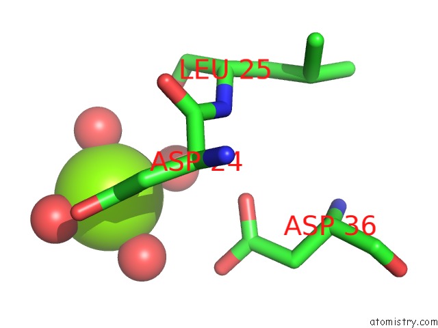
Magnesium binding site 5 out of 5 in 1kfg
Go back to
Magnesium binding site 5 out
of 5 in the The X-Ray Crystal Structure of CEL9G From Clostridium Cellulolyticum Complexed with A Thio-Oligosaccharide Inhibitor
Mono view
Stereo pair view
Mono view
Stereo pair view
A full contact list of Magnesium with other atoms in the Mg binding
site number 5 of The X-Ray Crystal Structure of CEL9G From Clostridium Cellulolyticum Complexed with A Thio-Oligosaccharide Inhibitor within 5.0Å range:
|
Reference:
D.Mandelman,
A.Belaich,
J.-P.Belaich,
N.Aghajari,
H.Driguez,
R.Haser.
The X-Ray Crystal Structure of the Multidomain Endoglucanase CEL9G From Clostridium Cellulolyticum Complexed with Natural and Synthetic Cello-Olligosaccharides J.Bacteriol. V. 185 4127 2003.
ISSN: ISSN 0021-9193
PubMed: 12837787
DOI: 10.1128/JB.185.14.4127-4135.2003
Page generated: Tue Aug 13 07:42:04 2024
ISSN: ISSN 0021-9193
PubMed: 12837787
DOI: 10.1128/JB.185.14.4127-4135.2003
Last articles
Zn in 9J0NZn in 9J0O
Zn in 9J0P
Zn in 9FJX
Zn in 9EKB
Zn in 9C0F
Zn in 9CAH
Zn in 9CH0
Zn in 9CH3
Zn in 9CH1