Magnesium »
PDB 1k9w-1kk3 »
1kfs »
Magnesium in PDB 1kfs: Dna Polymerase I Klenow Fragment (E.C.2.7.7.7) Mutant/Dna Complex
Enzymatic activity of Dna Polymerase I Klenow Fragment (E.C.2.7.7.7) Mutant/Dna Complex
All present enzymatic activity of Dna Polymerase I Klenow Fragment (E.C.2.7.7.7) Mutant/Dna Complex:
2.7.7.7;
2.7.7.7;
Protein crystallography data
The structure of Dna Polymerase I Klenow Fragment (E.C.2.7.7.7) Mutant/Dna Complex, PDB code: 1kfs
was solved by
C.A.Brautigam,
T.A.Steitz,
with X-Ray Crystallography technique. A brief refinement statistics is given in the table below:
| Resolution Low / High (Å) | 20.00 / 2.10 |
| Space group | P 43 |
| Cell size a, b, c (Å), α, β, γ (°) | 101.700, 101.700, 85.800, 90.00, 90.00, 90.00 |
| R / Rfree (%) | 19.5 / 21.9 |
Other elements in 1kfs:
The structure of Dna Polymerase I Klenow Fragment (E.C.2.7.7.7) Mutant/Dna Complex also contains other interesting chemical elements:
| Zinc | (Zn) | 3 atoms |
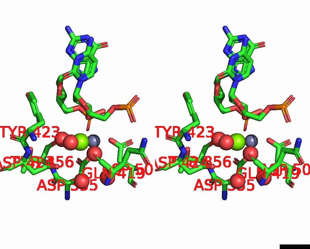
Magnesium Binding Sites:
The binding sites of Magnesium atom in the Dna Polymerase I Klenow Fragment (E.C.2.7.7.7) Mutant/Dna Complex
(pdb code 1kfs). This binding sites where shown within
5.0 Angstroms radius around Magnesium atom.
In total only one binding site of Magnesium was determined in the Dna Polymerase I Klenow Fragment (E.C.2.7.7.7) Mutant/Dna Complex, PDB code: 1kfs:
In total only one binding site of Magnesium was determined in the Dna Polymerase I Klenow Fragment (E.C.2.7.7.7) Mutant/Dna Complex, PDB code: 1kfs:
Magnesium binding site 1 out of 1 in 1kfs
Go back to
Magnesium binding site 1 out
of 1 in the Dna Polymerase I Klenow Fragment (E.C.2.7.7.7) Mutant/Dna Complex

Mono view
Stereo pair view
Mono view
Stereo pair view
A full contact list of Magnesium with other atoms in the Mg binding
site number 1 of Dna Polymerase I Klenow Fragment (E.C.2.7.7.7) Mutant/Dna Complex within 5.0Å range:
|
Reference:
C.A.Brautigam,
T.A.Steitz.
Structural Principles For the Inhibition of the 3'-5' Exonuclease Activity of Escherichia Coli Dna Polymerase I By Phosphorothioates. J.Mol.Biol. V. 277 363 1998.
ISSN: ISSN 0022-2836
PubMed: 9514742
DOI: 10.1006/JMBI.1997.1586
Page generated: Sun Aug 10 00:08:40 2025
ISSN: ISSN 0022-2836
PubMed: 9514742
DOI: 10.1006/JMBI.1997.1586
Last articles
Mg in 1YQWMg in 1YQT
Mg in 1YQ9
Mg in 1YQ7
Mg in 1YQ2
Mg in 1YNS
Mg in 1YNO
Mg in 1YMV
Mg in 1YMQ
Mg in 1YM3