Magnesium »
PDB 1kk7-1l3p »
1l3j »
Magnesium in PDB 1l3j: Crystal Structure of Oxalate Decarboxylase Formate Complex
Protein crystallography data
The structure of Crystal Structure of Oxalate Decarboxylase Formate Complex, PDB code: 1l3j
was solved by
R.Anand,
P.C.Dorrestein,
C.Kinsland,
T.P.Begley,
S.E.Ealick,
with X-Ray Crystallography technique. A brief refinement statistics is given in the table below:
| Resolution Low / High (Å) | 40.72 / 1.90 |
| Space group | H 3 2 |
| Cell size a, b, c (Å), α, β, γ (°) | 154.800, 154.800, 122.160, 90.00, 90.00, 120.00 |
| R / Rfree (%) | 16.9 / 19.3 |
Other elements in 1l3j:
The structure of Crystal Structure of Oxalate Decarboxylase Formate Complex also contains other interesting chemical elements:
| Manganese | (Mn) | 2 atoms |
Magnesium Binding Sites:
The binding sites of Magnesium atom in the Crystal Structure of Oxalate Decarboxylase Formate Complex
(pdb code 1l3j). This binding sites where shown within
5.0 Angstroms radius around Magnesium atom.
In total only one binding site of Magnesium was determined in the Crystal Structure of Oxalate Decarboxylase Formate Complex, PDB code: 1l3j:
In total only one binding site of Magnesium was determined in the Crystal Structure of Oxalate Decarboxylase Formate Complex, PDB code: 1l3j:
Magnesium binding site 1 out of 1 in 1l3j
Go back to
Magnesium binding site 1 out
of 1 in the Crystal Structure of Oxalate Decarboxylase Formate Complex
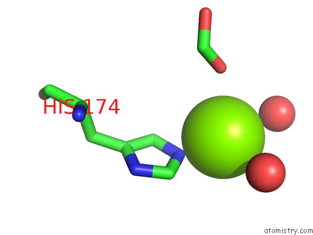
Mono view
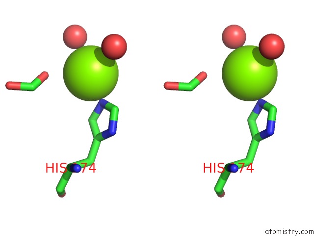
Stereo pair view
Mono view
Stereo pair view
A full contact list of Magnesium with other atoms in the Mg binding
site number 1 of Crystal Structure of Oxalate Decarboxylase Formate Complex within 5.0Å range:
|
Reference:
R.Anand,
P.C.Dorrestein,
C.Kinsland,
T.P.Begley,
S.E.Ealick.
Structure of Oxalate Decarboxylase From Bacillus Subtilis at 1.75 A Resolution. Biochemistry V. 41 7659 2002.
ISSN: ISSN 0006-2960
PubMed: 12056897
DOI: 10.1021/BI0200965
Page generated: Tue Aug 13 08:13:10 2024
ISSN: ISSN 0006-2960
PubMed: 12056897
DOI: 10.1021/BI0200965
Last articles
Zn in 9MJ5Zn in 9HNW
Zn in 9G0L
Zn in 9FNE
Zn in 9DZN
Zn in 9E0I
Zn in 9D32
Zn in 9DAK
Zn in 8ZXC
Zn in 8ZUF