Magnesium »
PDB 1l3r-1lny »
1l3t »
Magnesium in PDB 1l3t: Crystal Structure of Bacillus Dna Polymerase I Fragment Product Complex with 10 Base Pairs of Duplex Dna Following Addition of A Single Dttp Residue
Enzymatic activity of Crystal Structure of Bacillus Dna Polymerase I Fragment Product Complex with 10 Base Pairs of Duplex Dna Following Addition of A Single Dttp Residue
All present enzymatic activity of Crystal Structure of Bacillus Dna Polymerase I Fragment Product Complex with 10 Base Pairs of Duplex Dna Following Addition of A Single Dttp Residue:
2.7.7.7;
2.7.7.7;
Protein crystallography data
The structure of Crystal Structure of Bacillus Dna Polymerase I Fragment Product Complex with 10 Base Pairs of Duplex Dna Following Addition of A Single Dttp Residue, PDB code: 1l3t
was solved by
S.J.Johnson,
J.S.Taylor,
L.S.Beese,
with X-Ray Crystallography technique. A brief refinement statistics is given in the table below:
| Resolution Low / High (Å) | 35.06 / 1.70 |
| Space group | P 21 21 21 |
| Cell size a, b, c (Å), α, β, γ (°) | 87.530, 93.356, 106.229, 90.00, 90.00, 90.00 |
| R / Rfree (%) | 20.6 / 23.1 |
Magnesium Binding Sites:
The binding sites of Magnesium atom in the Crystal Structure of Bacillus Dna Polymerase I Fragment Product Complex with 10 Base Pairs of Duplex Dna Following Addition of A Single Dttp Residue
(pdb code 1l3t). This binding sites where shown within
5.0 Angstroms radius around Magnesium atom.
In total only one binding site of Magnesium was determined in the Crystal Structure of Bacillus Dna Polymerase I Fragment Product Complex with 10 Base Pairs of Duplex Dna Following Addition of A Single Dttp Residue, PDB code: 1l3t:
In total only one binding site of Magnesium was determined in the Crystal Structure of Bacillus Dna Polymerase I Fragment Product Complex with 10 Base Pairs of Duplex Dna Following Addition of A Single Dttp Residue, PDB code: 1l3t:
Magnesium binding site 1 out of 1 in 1l3t
Go back to
Magnesium binding site 1 out
of 1 in the Crystal Structure of Bacillus Dna Polymerase I Fragment Product Complex with 10 Base Pairs of Duplex Dna Following Addition of A Single Dttp Residue
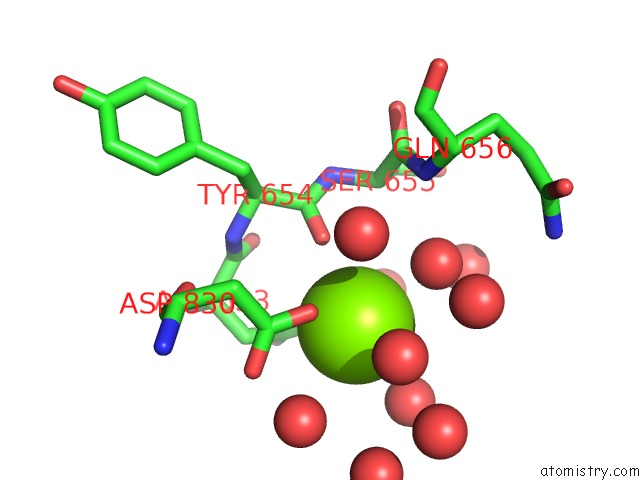
Mono view
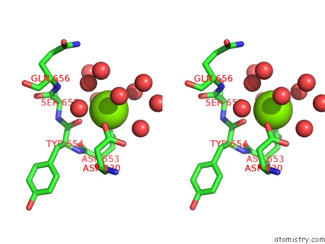
Stereo pair view
Mono view
Stereo pair view
A full contact list of Magnesium with other atoms in the Mg binding
site number 1 of Crystal Structure of Bacillus Dna Polymerase I Fragment Product Complex with 10 Base Pairs of Duplex Dna Following Addition of A Single Dttp Residue within 5.0Å range:
|
Reference:
S.J.Johnson,
J.S.Taylor,
L.S.Beese.
Processive Dna Synthesis Observed in A Polymerase Crystal Suggests A Mechanism For the Prevention of Frameshift Mutations Proc.Natl.Acad.Sci.Usa V. 100 3895 2003.
ISSN: ISSN 0027-8424
PubMed: 12649320
DOI: 10.1073/PNAS.0630532100
Page generated: Sun Aug 10 00:37:34 2025
ISSN: ISSN 0027-8424
PubMed: 12649320
DOI: 10.1073/PNAS.0630532100
Last articles
Mg in 4DPGMg in 4DQP
Mg in 4DQQ
Mg in 4DPM
Mg in 4DPV
Mg in 4DQI
Mg in 4DOB
Mg in 4DOC
Mg in 4DMZ
Mg in 4DOA