Magnesium »
PDB 1l3r-1lny »
1lij »
Magnesium in PDB 1lij: Structure of T. Gondii Adenosine Kinase Bound to Prodrug 2 7-Iodotubercidin and Amp-Pcp
Enzymatic activity of Structure of T. Gondii Adenosine Kinase Bound to Prodrug 2 7-Iodotubercidin and Amp-Pcp
All present enzymatic activity of Structure of T. Gondii Adenosine Kinase Bound to Prodrug 2 7-Iodotubercidin and Amp-Pcp:
2.7.1.20;
2.7.1.20;
Protein crystallography data
The structure of Structure of T. Gondii Adenosine Kinase Bound to Prodrug 2 7-Iodotubercidin and Amp-Pcp, PDB code: 1lij
was solved by
M.A.Schumacher,
D.M.Scott,
I.I.Mathews,
S.E.Ealick,
D.S.Roos,
B.Ullman,
R.G.Brennan,
with X-Ray Crystallography technique. A brief refinement statistics is given in the table below:
| Resolution Low / High (Å) | 10.00 / 1.86 |
| Space group | P 21 21 2 |
| Cell size a, b, c (Å), α, β, γ (°) | 167.700, 46.970, 44.090, 90.00, 90.00, 90.00 |
| R / Rfree (%) | n/a / n/a |
Other elements in 1lij:
The structure of Structure of T. Gondii Adenosine Kinase Bound to Prodrug 2 7-Iodotubercidin and Amp-Pcp also contains other interesting chemical elements:
| Iodine | (I) | 1 atom |
| Chlorine | (Cl) | 1 atom |
Magnesium Binding Sites:
The binding sites of Magnesium atom in the Structure of T. Gondii Adenosine Kinase Bound to Prodrug 2 7-Iodotubercidin and Amp-Pcp
(pdb code 1lij). This binding sites where shown within
5.0 Angstroms radius around Magnesium atom.
In total only one binding site of Magnesium was determined in the Structure of T. Gondii Adenosine Kinase Bound to Prodrug 2 7-Iodotubercidin and Amp-Pcp, PDB code: 1lij:
In total only one binding site of Magnesium was determined in the Structure of T. Gondii Adenosine Kinase Bound to Prodrug 2 7-Iodotubercidin and Amp-Pcp, PDB code: 1lij:
Magnesium binding site 1 out of 1 in 1lij
Go back to
Magnesium binding site 1 out
of 1 in the Structure of T. Gondii Adenosine Kinase Bound to Prodrug 2 7-Iodotubercidin and Amp-Pcp
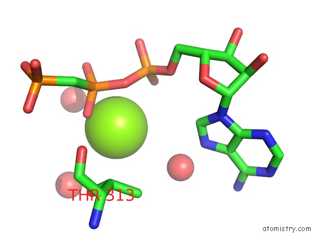
Mono view
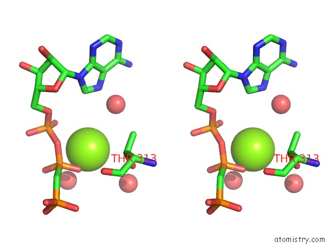
Stereo pair view
Mono view
Stereo pair view
A full contact list of Magnesium with other atoms in the Mg binding
site number 1 of Structure of T. Gondii Adenosine Kinase Bound to Prodrug 2 7-Iodotubercidin and Amp-Pcp within 5.0Å range:
|
Reference:
M.A.Schumacher,
D.M.Scott,
I.I.Mathews,
S.E.Ealick,
D.S.Roos,
B.Ullman,
R.G.Brennan.
Crystal Structures of Toxoplasma Gondii Adenosine Kinase Reveal A Novel Catalytic Mechanism and Prodrug Binding. J.Mol.Biol. V. 298 875 2000.
ISSN: ISSN 0022-2836
PubMed: 10801355
DOI: 10.1006/JMBI.2000.3753
Page generated: Tue Aug 13 08:31:17 2024
ISSN: ISSN 0022-2836
PubMed: 10801355
DOI: 10.1006/JMBI.2000.3753
Last articles
Zn in 9J0NZn in 9J0O
Zn in 9J0P
Zn in 9FJX
Zn in 9EKB
Zn in 9C0F
Zn in 9CAH
Zn in 9CH0
Zn in 9CH3
Zn in 9CH1