Magnesium »
PDB 1lnz-1mez »
1m3u »
Magnesium in PDB 1m3u: Crystal Structure of Ketopantoate Hydroxymethyltransferase Complexed the Product Ketopantoate
Enzymatic activity of Crystal Structure of Ketopantoate Hydroxymethyltransferase Complexed the Product Ketopantoate
All present enzymatic activity of Crystal Structure of Ketopantoate Hydroxymethyltransferase Complexed the Product Ketopantoate:
2.1.2.11;
2.1.2.11;
Protein crystallography data
The structure of Crystal Structure of Ketopantoate Hydroxymethyltransferase Complexed the Product Ketopantoate, PDB code: 1m3u
was solved by
F.Von Delft,
T.Inoue,
S.A.Saldanha,
H.H.Ottenhof,
V.Dhanaraj,
M.Witty,
C.Abell,
A.G.Smith,
T.L.Blundell,
with X-Ray Crystallography technique. A brief refinement statistics is given in the table below:
| Resolution Low / High (Å) | 100.00 / 1.80 |
| Space group | P 1 21 1 |
| Cell size a, b, c (Å), α, β, γ (°) | 86.074, 157.170, 100.181, 90.00, 97.44, 90.00 |
| R / Rfree (%) | 15.2 / 19.3 |
Magnesium Binding Sites:
The binding sites of Magnesium atom in the Crystal Structure of Ketopantoate Hydroxymethyltransferase Complexed the Product Ketopantoate
(pdb code 1m3u). This binding sites where shown within
5.0 Angstroms radius around Magnesium atom.
In total 10 binding sites of Magnesium where determined in the Crystal Structure of Ketopantoate Hydroxymethyltransferase Complexed the Product Ketopantoate, PDB code: 1m3u:
Jump to Magnesium binding site number: 1; 2; 3; 4; 5; 6; 7; 8; 9; 10;
In total 10 binding sites of Magnesium where determined in the Crystal Structure of Ketopantoate Hydroxymethyltransferase Complexed the Product Ketopantoate, PDB code: 1m3u:
Jump to Magnesium binding site number: 1; 2; 3; 4; 5; 6; 7; 8; 9; 10;
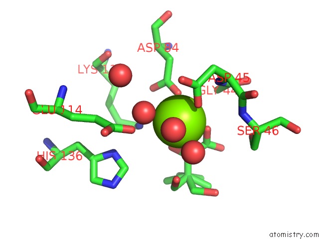
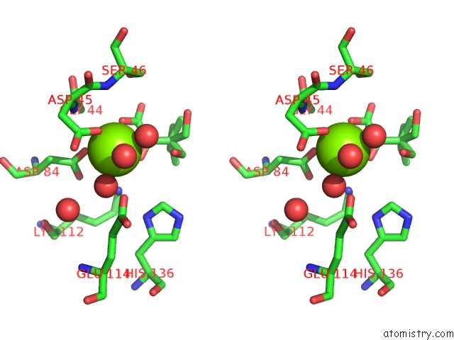
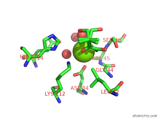
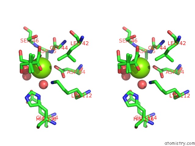
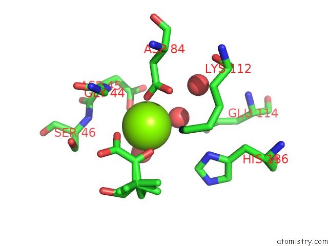
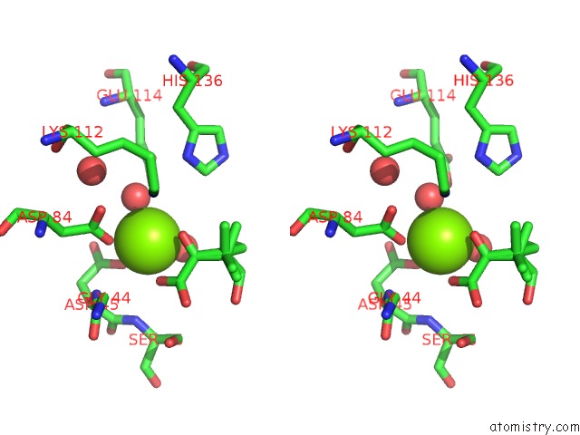
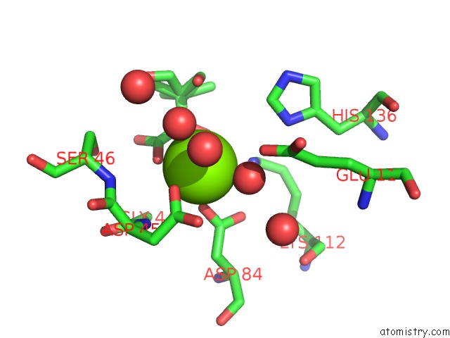
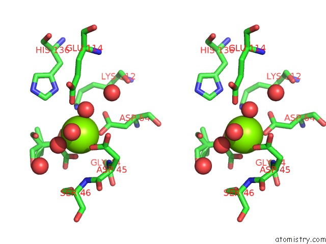
Magnesium binding site 1 out of 10 in 1m3u
Go back to
Magnesium binding site 1 out
of 10 in the Crystal Structure of Ketopantoate Hydroxymethyltransferase Complexed the Product Ketopantoate
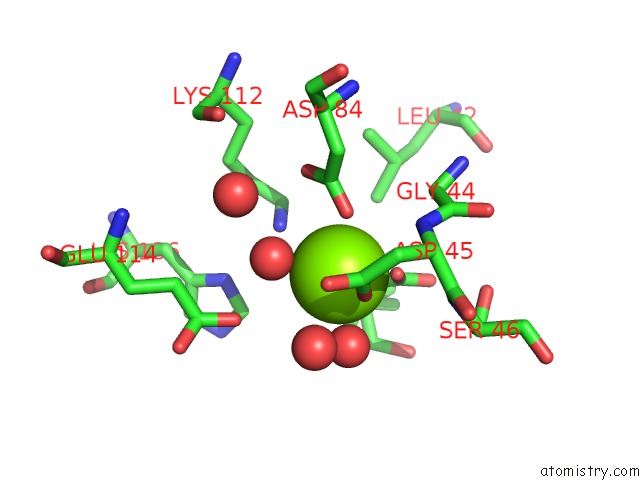
Mono view
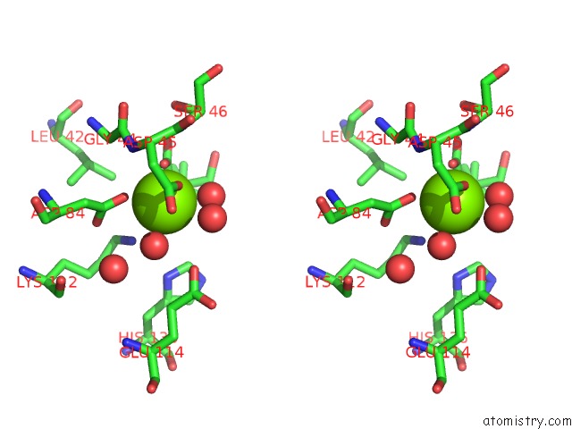
Stereo pair view
Mono view
Stereo pair view
A full contact list of Magnesium with other atoms in the Mg binding
site number 1 of Crystal Structure of Ketopantoate Hydroxymethyltransferase Complexed the Product Ketopantoate within 5.0Å range:
|
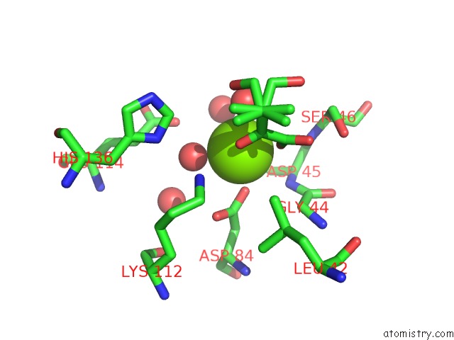
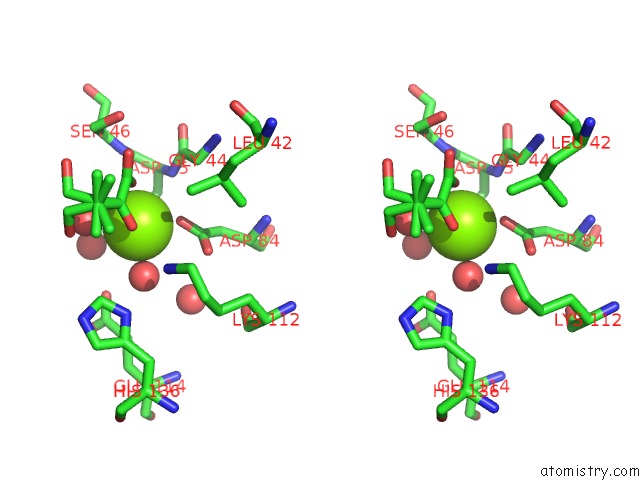
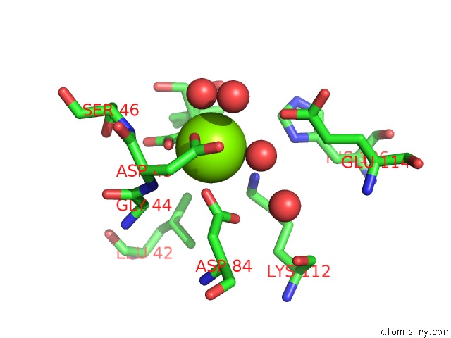
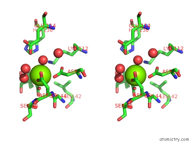
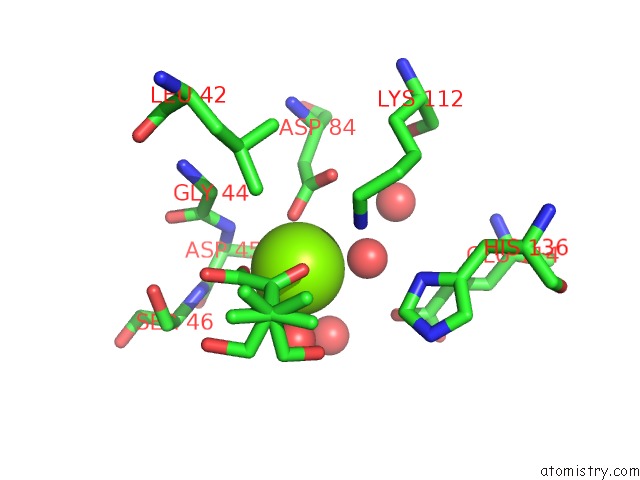
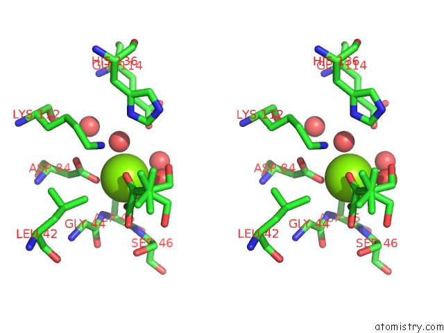
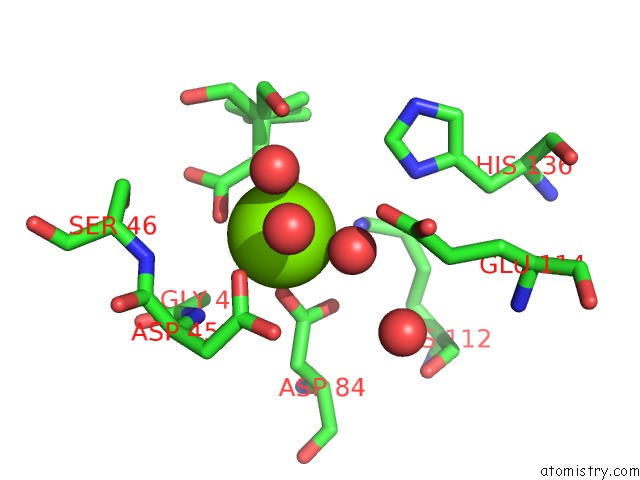
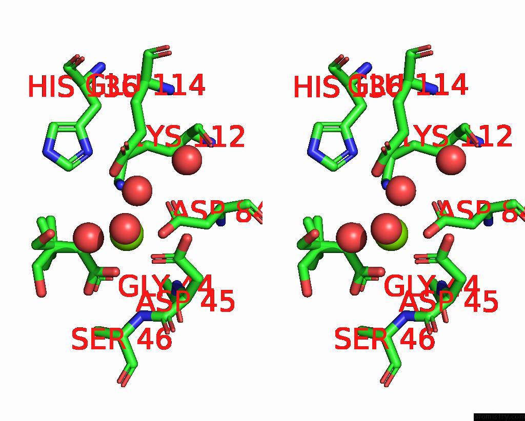
Magnesium binding site 2 out of 10 in 1m3u
Go back to
Magnesium binding site 2 out
of 10 in the Crystal Structure of Ketopantoate Hydroxymethyltransferase Complexed the Product Ketopantoate
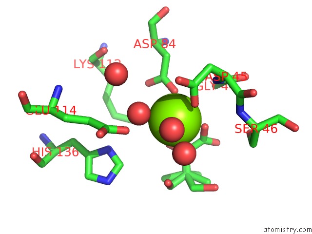
Mono view
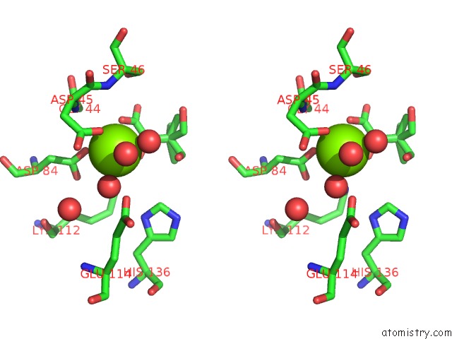
Stereo pair view
Mono view
Stereo pair view
A full contact list of Magnesium with other atoms in the Mg binding
site number 2 of Crystal Structure of Ketopantoate Hydroxymethyltransferase Complexed the Product Ketopantoate within 5.0Å range:
|
Magnesium binding site 3 out of 10 in 1m3u
Go back to
Magnesium binding site 3 out
of 10 in the Crystal Structure of Ketopantoate Hydroxymethyltransferase Complexed the Product Ketopantoate
Mono view
Stereo pair view
Mono view
Stereo pair view
A full contact list of Magnesium with other atoms in the Mg binding
site number 3 of Crystal Structure of Ketopantoate Hydroxymethyltransferase Complexed the Product Ketopantoate within 5.0Å range:
|
Magnesium binding site 4 out of 10 in 1m3u
Go back to
Magnesium binding site 4 out
of 10 in the Crystal Structure of Ketopantoate Hydroxymethyltransferase Complexed the Product Ketopantoate
Mono view
Stereo pair view
Mono view
Stereo pair view
A full contact list of Magnesium with other atoms in the Mg binding
site number 4 of Crystal Structure of Ketopantoate Hydroxymethyltransferase Complexed the Product Ketopantoate within 5.0Å range:
|
Magnesium binding site 5 out of 10 in 1m3u
Go back to
Magnesium binding site 5 out
of 10 in the Crystal Structure of Ketopantoate Hydroxymethyltransferase Complexed the Product Ketopantoate
Mono view
Stereo pair view
Mono view
Stereo pair view
A full contact list of Magnesium with other atoms in the Mg binding
site number 5 of Crystal Structure of Ketopantoate Hydroxymethyltransferase Complexed the Product Ketopantoate within 5.0Å range:
|
Magnesium binding site 6 out of 10 in 1m3u
Go back to
Magnesium binding site 6 out
of 10 in the Crystal Structure of Ketopantoate Hydroxymethyltransferase Complexed the Product Ketopantoate
Mono view
Stereo pair view
Mono view
Stereo pair view
A full contact list of Magnesium with other atoms in the Mg binding
site number 6 of Crystal Structure of Ketopantoate Hydroxymethyltransferase Complexed the Product Ketopantoate within 5.0Å range:
|
Magnesium binding site 7 out of 10 in 1m3u
Go back to
Magnesium binding site 7 out
of 10 in the Crystal Structure of Ketopantoate Hydroxymethyltransferase Complexed the Product Ketopantoate
Mono view
Stereo pair view
Mono view
Stereo pair view
A full contact list of Magnesium with other atoms in the Mg binding
site number 7 of Crystal Structure of Ketopantoate Hydroxymethyltransferase Complexed the Product Ketopantoate within 5.0Å range:
|
Magnesium binding site 8 out of 10 in 1m3u
Go back to
Magnesium binding site 8 out
of 10 in the Crystal Structure of Ketopantoate Hydroxymethyltransferase Complexed the Product Ketopantoate
Mono view
Stereo pair view
Mono view
Stereo pair view
A full contact list of Magnesium with other atoms in the Mg binding
site number 8 of Crystal Structure of Ketopantoate Hydroxymethyltransferase Complexed the Product Ketopantoate within 5.0Å range:
|
Magnesium binding site 9 out of 10 in 1m3u
Go back to
Magnesium binding site 9 out
of 10 in the Crystal Structure of Ketopantoate Hydroxymethyltransferase Complexed the Product Ketopantoate
Mono view
Stereo pair view
Mono view
Stereo pair view
A full contact list of Magnesium with other atoms in the Mg binding
site number 9 of Crystal Structure of Ketopantoate Hydroxymethyltransferase Complexed the Product Ketopantoate within 5.0Å range:
|
Magnesium binding site 10 out of 10 in 1m3u
Go back to
Magnesium binding site 10 out
of 10 in the Crystal Structure of Ketopantoate Hydroxymethyltransferase Complexed the Product Ketopantoate
Mono view
Stereo pair view
Mono view
Stereo pair view
A full contact list of Magnesium with other atoms in the Mg binding
site number 10 of Crystal Structure of Ketopantoate Hydroxymethyltransferase Complexed the Product Ketopantoate within 5.0Å range:
|
Reference:
F.Von Delft,
T.Inoue,
S.A.Saldanha,
H.H.Ottenhof,
F.Schmitzberger,
L.M.Birch,
V.Dhanaraj,
M.Witty,
A.G.Smith,
T.L.Blundell,
C.Abell.
Structure of E. Coli Ketopantoate Hydroxymethyl Transferase Complexed with Ketopantoate and Mg(2+), Solved By Locating 160 Selenomethionine Sites. Structure V. 11 985 2003.
ISSN: ISSN 0969-2126
PubMed: 12906829
DOI: 10.1016/S0969-2126(03)00158-8
Page generated: Sun Aug 10 00:45:49 2025
ISSN: ISSN 0969-2126
PubMed: 12906829
DOI: 10.1016/S0969-2126(03)00158-8
Last articles
Mg in 4F86Mg in 4F9A
Mg in 4F99
Mg in 4F93
Mg in 4F8B
Mg in 4F8E
Mg in 4F71
Mg in 4F72
Mg in 4F6X
Mg in 4F61