Magnesium »
PDB 1lnz-1mez »
1ma9 »
Magnesium in PDB 1ma9: Crystal Structure of the Complex of Human Vitamin D Binding Protein and Rabbit Muscle Actin
Protein crystallography data
The structure of Crystal Structure of the Complex of Human Vitamin D Binding Protein and Rabbit Muscle Actin, PDB code: 1ma9
was solved by
C.Verboven,
I.Bogaerts,
E.Waelkens,
A.Rabijns,
H.Van Baelen,
R.Bouillon,
C.De Ranter,
with X-Ray Crystallography technique. A brief refinement statistics is given in the table below:
| Resolution Low / High (Å) | 19.91 / 2.40 |
| Space group | P 1 21 1 |
| Cell size a, b, c (Å), α, β, γ (°) | 74.440, 74.900, 88.020, 90.00, 110.19, 90.00 |
| R / Rfree (%) | 20 / 25 |
Magnesium Binding Sites:
The binding sites of Magnesium atom in the Crystal Structure of the Complex of Human Vitamin D Binding Protein and Rabbit Muscle Actin
(pdb code 1ma9). This binding sites where shown within
5.0 Angstroms radius around Magnesium atom.
In total only one binding site of Magnesium was determined in the Crystal Structure of the Complex of Human Vitamin D Binding Protein and Rabbit Muscle Actin, PDB code: 1ma9:
In total only one binding site of Magnesium was determined in the Crystal Structure of the Complex of Human Vitamin D Binding Protein and Rabbit Muscle Actin, PDB code: 1ma9:
Magnesium binding site 1 out of 1 in 1ma9
Go back to
Magnesium binding site 1 out
of 1 in the Crystal Structure of the Complex of Human Vitamin D Binding Protein and Rabbit Muscle Actin
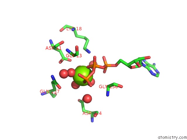
Mono view
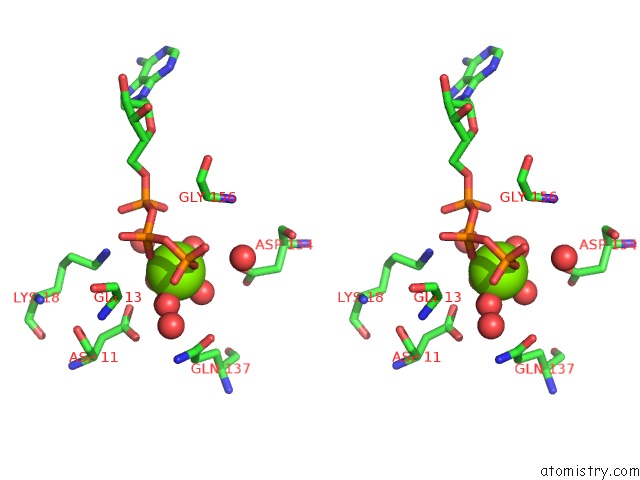
Stereo pair view
Mono view
Stereo pair view
A full contact list of Magnesium with other atoms in the Mg binding
site number 1 of Crystal Structure of the Complex of Human Vitamin D Binding Protein and Rabbit Muscle Actin within 5.0Å range:
|
Reference:
C.Verboven,
I.Bogaerts,
E.Waelkens,
A.Rabijns,
H.Van Baelen,
R.Bouillon,
C.De Ranter.
Actin-Dbp: the Perfect Structural Fit? Acta Crystallogr.,Sect.D V. 59 263 2003.
ISSN: ISSN 0907-4449
PubMed: 12554937
DOI: 10.1107/S0907444902021455
Page generated: Sun Aug 10 00:48:05 2025
ISSN: ISSN 0907-4449
PubMed: 12554937
DOI: 10.1107/S0907444902021455
Last articles
Mg in 5D9BMg in 5D8N
Mg in 5D92
Mg in 5D91
Mg in 5D8G
Mg in 5D7R
Mg in 5D87
Mg in 5D5L
Mg in 5D86
Mg in 5D7D