Magnesium »
PDB 1mf0-1muh »
1mh9 »
Magnesium in PDB 1mh9: Crystal Structure Analysis of Deoxyribonucleotidase
Enzymatic activity of Crystal Structure Analysis of Deoxyribonucleotidase
All present enzymatic activity of Crystal Structure Analysis of Deoxyribonucleotidase:
3.1.3.5;
3.1.3.5;
Protein crystallography data
The structure of Crystal Structure Analysis of Deoxyribonucleotidase, PDB code: 1mh9
was solved by
A.Rinaldo-Matthis,
C.Rampazzo,
P.Reichard,
V.Bianchi,
P.Nordlund,
with X-Ray Crystallography technique. A brief refinement statistics is given in the table below:
| Resolution Low / High (Å) | 19.64 / 1.80 |
| Space group | P 43 21 2 |
| Cell size a, b, c (Å), α, β, γ (°) | 73.965, 73.965, 105.932, 90.00, 90.00, 90.00 |
| R / Rfree (%) | 15.9 / 19.1 |
Magnesium Binding Sites:
The binding sites of Magnesium atom in the Crystal Structure Analysis of Deoxyribonucleotidase
(pdb code 1mh9). This binding sites where shown within
5.0 Angstroms radius around Magnesium atom.
In total only one binding site of Magnesium was determined in the Crystal Structure Analysis of Deoxyribonucleotidase, PDB code: 1mh9:
In total only one binding site of Magnesium was determined in the Crystal Structure Analysis of Deoxyribonucleotidase, PDB code: 1mh9:
Magnesium binding site 1 out of 1 in 1mh9
Go back to
Magnesium binding site 1 out
of 1 in the Crystal Structure Analysis of Deoxyribonucleotidase
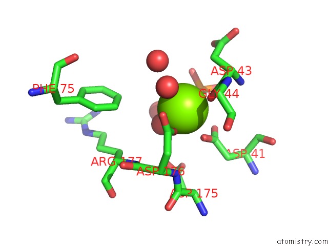
Mono view
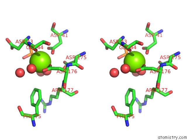
Stereo pair view
Mono view
Stereo pair view
A full contact list of Magnesium with other atoms in the Mg binding
site number 1 of Crystal Structure Analysis of Deoxyribonucleotidase within 5.0Å range:
|
Reference:
A.Rinaldo-Matthis,
C.Rampazzo,
P.Reichard,
V.Bianchi,
P.Nordlund.
Crystal Structure of A Human Mitochondrial Deoxyribonucleotidase. Nat.Struct.Biol. V. 9 779 2002.
ISSN: ISSN 1072-8368
PubMed: 12352955
DOI: 10.1038/NSB846
Page generated: Sun Aug 10 01:04:45 2025
ISSN: ISSN 1072-8368
PubMed: 12352955
DOI: 10.1038/NSB846
Last articles
Mg in 5D9BMg in 5D8N
Mg in 5D92
Mg in 5D91
Mg in 5D8G
Mg in 5D7R
Mg in 5D87
Mg in 5D5L
Mg in 5D86
Mg in 5D7D