Magnesium »
PDB 1mf0-1muh »
1mma »
Magnesium in PDB 1mma: X-Ray Structures of the Mgadp, Mgatpgammas, and Mgamppnp Complexes of the Dictyostelium Discoideum Myosin Motor Domain
Enzymatic activity of X-Ray Structures of the Mgadp, Mgatpgammas, and Mgamppnp Complexes of the Dictyostelium Discoideum Myosin Motor Domain
All present enzymatic activity of X-Ray Structures of the Mgadp, Mgatpgammas, and Mgamppnp Complexes of the Dictyostelium Discoideum Myosin Motor Domain:
3.6.1.32;
3.6.1.32;
Protein crystallography data
The structure of X-Ray Structures of the Mgadp, Mgatpgammas, and Mgamppnp Complexes of the Dictyostelium Discoideum Myosin Motor Domain, PDB code: 1mma
was solved by
A.M.Gulick,
C.B.Bauer,
J.B.Thoden,
I.Rayment,
with X-Ray Crystallography technique. A brief refinement statistics is given in the table below:
| Resolution Low / High (Å) | 20.00 / 2.10 |
| Space group | P 21 21 2 |
| Cell size a, b, c (Å), α, β, γ (°) | 103.600, 179.000, 53.900, 90.00, 90.00, 90.00 |
| R / Rfree (%) | 21.9 / n/a |
Magnesium Binding Sites:
The binding sites of Magnesium atom in the X-Ray Structures of the Mgadp, Mgatpgammas, and Mgamppnp Complexes of the Dictyostelium Discoideum Myosin Motor Domain
(pdb code 1mma). This binding sites where shown within
5.0 Angstroms radius around Magnesium atom.
In total only one binding site of Magnesium was determined in the X-Ray Structures of the Mgadp, Mgatpgammas, and Mgamppnp Complexes of the Dictyostelium Discoideum Myosin Motor Domain, PDB code: 1mma:
In total only one binding site of Magnesium was determined in the X-Ray Structures of the Mgadp, Mgatpgammas, and Mgamppnp Complexes of the Dictyostelium Discoideum Myosin Motor Domain, PDB code: 1mma:
Magnesium binding site 1 out of 1 in 1mma
Go back to
Magnesium binding site 1 out
of 1 in the X-Ray Structures of the Mgadp, Mgatpgammas, and Mgamppnp Complexes of the Dictyostelium Discoideum Myosin Motor Domain
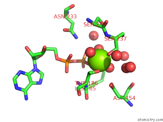
Mono view
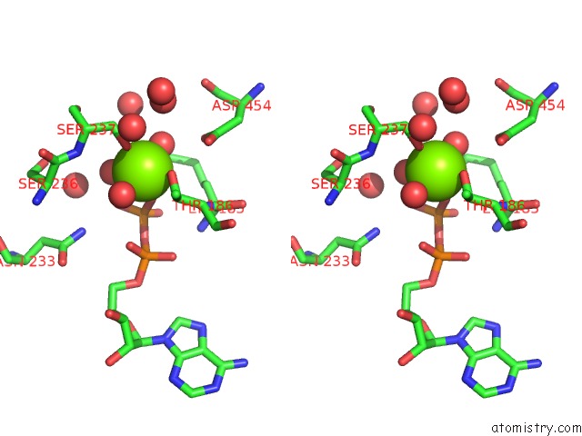
Stereo pair view
Mono view
Stereo pair view
A full contact list of Magnesium with other atoms in the Mg binding
site number 1 of X-Ray Structures of the Mgadp, Mgatpgammas, and Mgamppnp Complexes of the Dictyostelium Discoideum Myosin Motor Domain within 5.0Å range:
|
Reference:
A.M.Gulick,
C.B.Bauer,
J.B.Thoden,
I.Rayment.
X-Ray Structures of the Mgadp, Mgatpgammas, and Mgamppnp Complexes of the Dictyostelium Discoideum Myosin Motor Domain. Biochemistry V. 36 11619 1997.
ISSN: ISSN 0006-2960
PubMed: 9305951
DOI: 10.1021/BI9712596
Page generated: Sun Aug 10 01:06:21 2025
ISSN: ISSN 0006-2960
PubMed: 9305951
DOI: 10.1021/BI9712596
Last articles
Mg in 4DPGMg in 4DQP
Mg in 4DQQ
Mg in 4DPM
Mg in 4DPV
Mg in 4DQI
Mg in 4DOB
Mg in 4DOC
Mg in 4DMZ
Mg in 4DOA