Magnesium »
PDB 1mum-1n6i »
1mum »
Magnesium in PDB 1mum: Structure of the 2-Methylisocitrate Lyase (Prpb) From Escherichia Coli
Enzymatic activity of Structure of the 2-Methylisocitrate Lyase (Prpb) From Escherichia Coli
All present enzymatic activity of Structure of the 2-Methylisocitrate Lyase (Prpb) From Escherichia Coli:
4.1.3.30;
4.1.3.30;
Protein crystallography data
The structure of Structure of the 2-Methylisocitrate Lyase (Prpb) From Escherichia Coli, PDB code: 1mum
was solved by
C.Grimm,
K.Reuter,
with X-Ray Crystallography technique. A brief refinement statistics is given in the table below:
| Resolution Low / High (Å) | 29.35 / 1.90 |
| Space group | P 32 2 1 |
| Cell size a, b, c (Å), α, β, γ (°) | 82.895, 82.895, 166.250, 90.00, 90.00, 120.00 |
| R / Rfree (%) | 21.3 / 23.4 |
Magnesium Binding Sites:
The binding sites of Magnesium atom in the Structure of the 2-Methylisocitrate Lyase (Prpb) From Escherichia Coli
(pdb code 1mum). This binding sites where shown within
5.0 Angstroms radius around Magnesium atom.
In total 2 binding sites of Magnesium where determined in the Structure of the 2-Methylisocitrate Lyase (Prpb) From Escherichia Coli, PDB code: 1mum:
Jump to Magnesium binding site number: 1; 2;
In total 2 binding sites of Magnesium where determined in the Structure of the 2-Methylisocitrate Lyase (Prpb) From Escherichia Coli, PDB code: 1mum:
Jump to Magnesium binding site number: 1; 2;
Magnesium binding site 1 out of 2 in 1mum
Go back to
Magnesium binding site 1 out
of 2 in the Structure of the 2-Methylisocitrate Lyase (Prpb) From Escherichia Coli
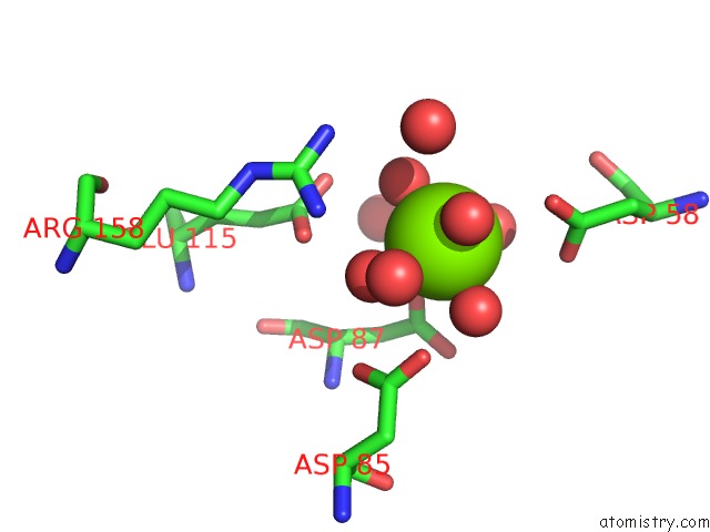
Mono view
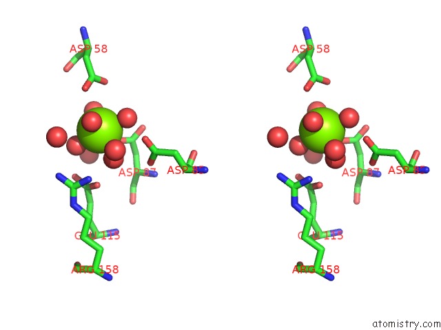
Stereo pair view
Mono view
Stereo pair view
A full contact list of Magnesium with other atoms in the Mg binding
site number 1 of Structure of the 2-Methylisocitrate Lyase (Prpb) From Escherichia Coli within 5.0Å range:
|
Magnesium binding site 2 out of 2 in 1mum
Go back to
Magnesium binding site 2 out
of 2 in the Structure of the 2-Methylisocitrate Lyase (Prpb) From Escherichia Coli
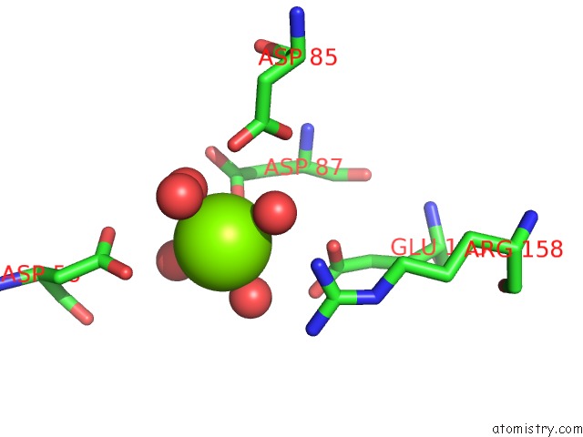
Mono view
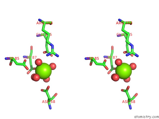
Stereo pair view
Mono view
Stereo pair view
A full contact list of Magnesium with other atoms in the Mg binding
site number 2 of Structure of the 2-Methylisocitrate Lyase (Prpb) From Escherichia Coli within 5.0Å range:
|
Reference:
C.Grimm,
A.Evers,
M.Brock,
C.Maerker,
G.Klebe,
W.Buckel,
K.Reuter.
Crystal Structure of 2-Methylisocitrate Lyase (Prpb) From Escherichia Coli and Modelling of Its Ligand Bound Active Centre. J.Mol.Biol. V. 328 609 2003.
ISSN: ISSN 0022-2836
PubMed: 12706720
DOI: 10.1016/S0022-2836(03)00358-9
Page generated: Sun Aug 10 01:13:40 2025
ISSN: ISSN 0022-2836
PubMed: 12706720
DOI: 10.1016/S0022-2836(03)00358-9
Last articles
Mg in 1WC1Mg in 1WBQ
Mg in 1WC5
Mg in 1WBD
Mg in 1WBB
Mg in 1WB9
Mg in 1WAX
Mg in 1WA5
Mg in 1W9L
Mg in 1W8Y