Magnesium »
PDB 1mum-1n6i »
1n1o »
Magnesium in PDB 1n1o: Crystal Structure of A B-Form Dna Duplex Containing (L)-Alpha- Threofuranosyl (3'-2') Nucleosides: A Four-Carbon Sugar Is Easily Accommodated Into the Backbone of Dna
Protein crystallography data
The structure of Crystal Structure of A B-Form Dna Duplex Containing (L)-Alpha- Threofuranosyl (3'-2') Nucleosides: A Four-Carbon Sugar Is Easily Accommodated Into the Backbone of Dna, PDB code: 1n1o
was solved by
C.J.Wilds,
Z.Wawrzak,
R.Krishnamurthy,
A.Eschenmoser,
M.Egli,
with X-Ray Crystallography technique. A brief refinement statistics is given in the table below:
| Resolution Low / High (Å) | 20.00 / 1.20 |
| Space group | P 21 21 21 |
| Cell size a, b, c (Å), α, β, γ (°) | 25.277, 40.080, 65.860, 90.00, 90.00, 90.00 |
| R / Rfree (%) | 16 / 20 |
Magnesium Binding Sites:
The binding sites of Magnesium atom in the Crystal Structure of A B-Form Dna Duplex Containing (L)-Alpha- Threofuranosyl (3'-2') Nucleosides: A Four-Carbon Sugar Is Easily Accommodated Into the Backbone of Dna
(pdb code 1n1o). This binding sites where shown within
5.0 Angstroms radius around Magnesium atom.
In total only one binding site of Magnesium was determined in the Crystal Structure of A B-Form Dna Duplex Containing (L)-Alpha- Threofuranosyl (3'-2') Nucleosides: A Four-Carbon Sugar Is Easily Accommodated Into the Backbone of Dna, PDB code: 1n1o:
In total only one binding site of Magnesium was determined in the Crystal Structure of A B-Form Dna Duplex Containing (L)-Alpha- Threofuranosyl (3'-2') Nucleosides: A Four-Carbon Sugar Is Easily Accommodated Into the Backbone of Dna, PDB code: 1n1o:
Magnesium binding site 1 out of 1 in 1n1o
Go back to
Magnesium binding site 1 out
of 1 in the Crystal Structure of A B-Form Dna Duplex Containing (L)-Alpha- Threofuranosyl (3'-2') Nucleosides: A Four-Carbon Sugar Is Easily Accommodated Into the Backbone of Dna
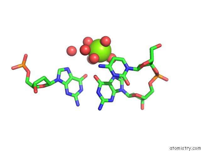
Mono view
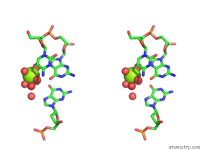
Stereo pair view
Mono view
Stereo pair view
A full contact list of Magnesium with other atoms in the Mg binding
site number 1 of Crystal Structure of A B-Form Dna Duplex Containing (L)-Alpha- Threofuranosyl (3'-2') Nucleosides: A Four-Carbon Sugar Is Easily Accommodated Into the Backbone of Dna within 5.0Å range:
|
Reference:
C.J.Wilds,
Z.Wawrzak,
R.Krishnamurthy,
A.Eschenmoser,
M.Egli.
Crystal Structure of A B-Form Dna Duplex Containing (L)-Alpha-Threofuranosyl (3'-->2') Nucleosides: A Four-Carbon Sugar Is Easily Accommodated Into the Backbone of Dna J.Am.Chem.Soc. V. 124 13716 2002.
ISSN: ISSN 0002-7863
PubMed: 12431101
DOI: 10.1021/JA0207807
Page generated: Tue Aug 13 09:18:31 2024
ISSN: ISSN 0002-7863
PubMed: 12431101
DOI: 10.1021/JA0207807
Last articles
Zn in 9J0NZn in 9J0O
Zn in 9J0P
Zn in 9FJX
Zn in 9EKB
Zn in 9C0F
Zn in 9CAH
Zn in 9CH0
Zn in 9CH3
Zn in 9CH1