Magnesium »
PDB 1nq6-1nzw »
1nr9 »
Magnesium in PDB 1nr9: Crystal Structure of Escherichia Coli 1262 (APC5008), Putative Isomerase
Protein crystallography data
The structure of Crystal Structure of Escherichia Coli 1262 (APC5008), Putative Isomerase, PDB code: 1nr9
was solved by
Y.Kim,
A.Joachimiak,
A.Edwards,
T.Skarina,
A.Savchenko,
Midwestcenter For Structural Genomics (Mcsg),
with X-Ray Crystallography technique. A brief refinement statistics is given in the table below:
| Resolution Low / High (Å) | 47.38 / 2.70 |
| Space group | P 1 21 1 |
| Cell size a, b, c (Å), α, β, γ (°) | 59.373, 83.546, 94.896, 90.00, 93.08, 90.00 |
| R / Rfree (%) | 20.6 / 27.5 |
Magnesium Binding Sites:
The binding sites of Magnesium atom in the Crystal Structure of Escherichia Coli 1262 (APC5008), Putative Isomerase
(pdb code 1nr9). This binding sites where shown within
5.0 Angstroms radius around Magnesium atom.
In total 4 binding sites of Magnesium where determined in the Crystal Structure of Escherichia Coli 1262 (APC5008), Putative Isomerase, PDB code: 1nr9:
Jump to Magnesium binding site number: 1; 2; 3; 4;
In total 4 binding sites of Magnesium where determined in the Crystal Structure of Escherichia Coli 1262 (APC5008), Putative Isomerase, PDB code: 1nr9:
Jump to Magnesium binding site number: 1; 2; 3; 4;
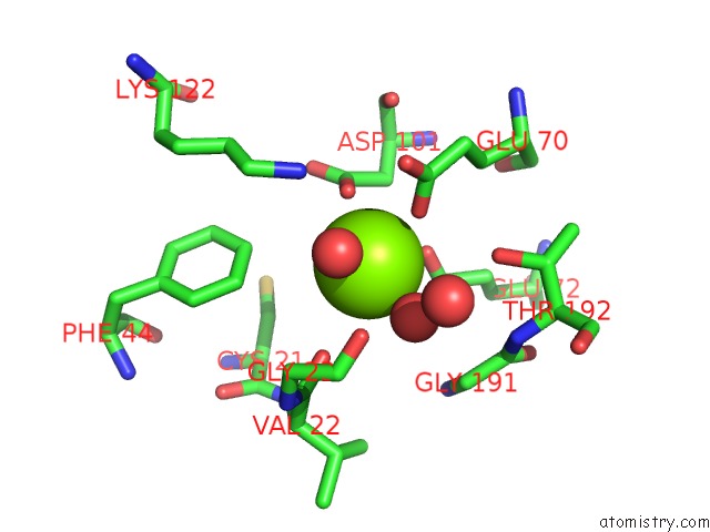
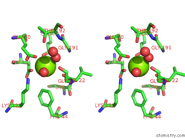
Magnesium binding site 1 out of 4 in 1nr9
Go back to
Magnesium binding site 1 out
of 4 in the Crystal Structure of Escherichia Coli 1262 (APC5008), Putative Isomerase
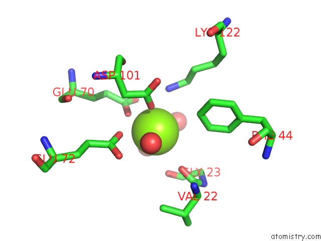
Mono view
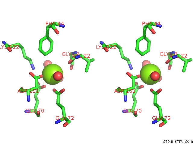
Stereo pair view
Mono view
Stereo pair view
A full contact list of Magnesium with other atoms in the Mg binding
site number 1 of Crystal Structure of Escherichia Coli 1262 (APC5008), Putative Isomerase within 5.0Å range:
|
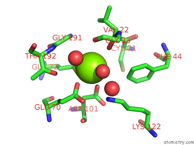
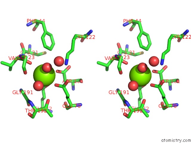
Magnesium binding site 2 out of 4 in 1nr9
Go back to
Magnesium binding site 2 out
of 4 in the Crystal Structure of Escherichia Coli 1262 (APC5008), Putative Isomerase
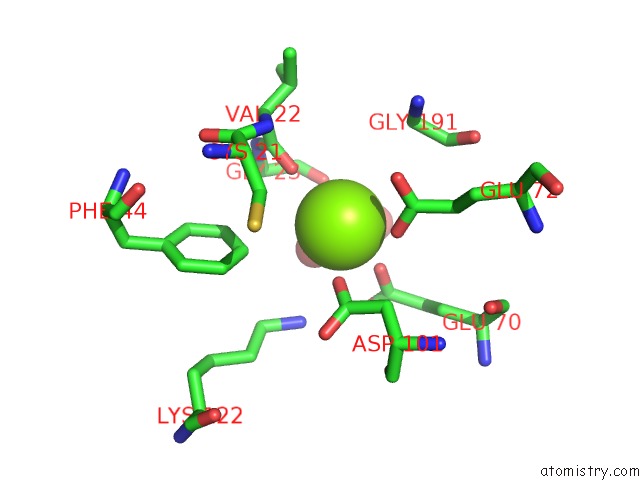
Mono view
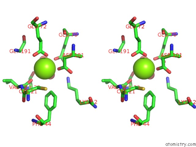
Stereo pair view
Mono view
Stereo pair view
A full contact list of Magnesium with other atoms in the Mg binding
site number 2 of Crystal Structure of Escherichia Coli 1262 (APC5008), Putative Isomerase within 5.0Å range:
|
Magnesium binding site 3 out of 4 in 1nr9
Go back to
Magnesium binding site 3 out
of 4 in the Crystal Structure of Escherichia Coli 1262 (APC5008), Putative Isomerase
Mono view
Stereo pair view
Mono view
Stereo pair view
A full contact list of Magnesium with other atoms in the Mg binding
site number 3 of Crystal Structure of Escherichia Coli 1262 (APC5008), Putative Isomerase within 5.0Å range:
|
Magnesium binding site 4 out of 4 in 1nr9
Go back to
Magnesium binding site 4 out
of 4 in the Crystal Structure of Escherichia Coli 1262 (APC5008), Putative Isomerase
Mono view
Stereo pair view
Mono view
Stereo pair view
A full contact list of Magnesium with other atoms in the Mg binding
site number 4 of Crystal Structure of Escherichia Coli 1262 (APC5008), Putative Isomerase within 5.0Å range:
|
Reference:
Y.Kim,
A.Joachimiak,
A.Edwards,
T.Skarina,
A.Savchenko.
Crystal Structure of Escherichia Coli Putative Isomerase EC1262 (APC5008) To Be Published.
Page generated: Tue Aug 13 10:21:52 2024
Last articles
Cl in 6BDTCl in 6BFJ
Cl in 6BEL
Cl in 6BB3
Cl in 6BEA
Cl in 6BE1
Cl in 6BDV
Cl in 6BB9
Cl in 6BCS
Cl in 6BBT