Magnesium »
PDB 1nq6-1nzw »
1nzi »
Magnesium in PDB 1nzi: Crystal Structure of the CUB1-Egf Interaction Domain of Complement Protease C1S
Enzymatic activity of Crystal Structure of the CUB1-Egf Interaction Domain of Complement Protease C1S
All present enzymatic activity of Crystal Structure of the CUB1-Egf Interaction Domain of Complement Protease C1S:
3.4.21.42;
3.4.21.42;
Protein crystallography data
The structure of Crystal Structure of the CUB1-Egf Interaction Domain of Complement Protease C1S, PDB code: 1nzi
was solved by
L.A.Gregory,
N.M.Thielens,
G.J.Arlaud,
J.C.Fontecilla-Camps,
C.Gaboriaud,
with X-Ray Crystallography technique. A brief refinement statistics is given in the table below:
| Resolution Low / High (Å) | 30.00 / 1.50 |
| Space group | P 1 |
| Cell size a, b, c (Å), α, β, γ (°) | 35.134, 47.499, 56.679, 87.74, 78.04, 75.67 |
| R / Rfree (%) | 21.6 / 23.4 |
Other elements in 1nzi:
The structure of Crystal Structure of the CUB1-Egf Interaction Domain of Complement Protease C1S also contains other interesting chemical elements:
| Calcium | (Ca) | 2 atoms |
Magnesium Binding Sites:
The binding sites of Magnesium atom in the Crystal Structure of the CUB1-Egf Interaction Domain of Complement Protease C1S
(pdb code 1nzi). This binding sites where shown within
5.0 Angstroms radius around Magnesium atom.
In total 2 binding sites of Magnesium where determined in the Crystal Structure of the CUB1-Egf Interaction Domain of Complement Protease C1S, PDB code: 1nzi:
Jump to Magnesium binding site number: 1; 2;
In total 2 binding sites of Magnesium where determined in the Crystal Structure of the CUB1-Egf Interaction Domain of Complement Protease C1S, PDB code: 1nzi:
Jump to Magnesium binding site number: 1; 2;
Magnesium binding site 1 out of 2 in 1nzi
Go back to
Magnesium binding site 1 out
of 2 in the Crystal Structure of the CUB1-Egf Interaction Domain of Complement Protease C1S
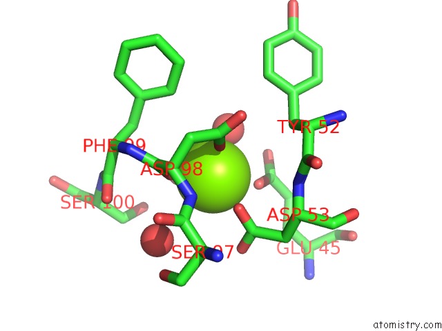
Mono view
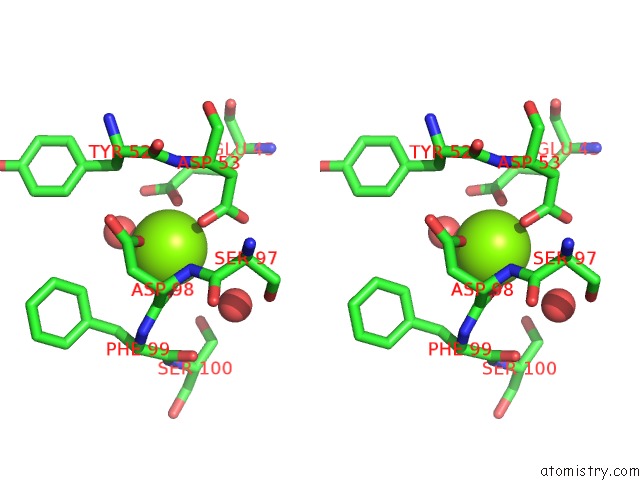
Stereo pair view
Mono view
Stereo pair view
A full contact list of Magnesium with other atoms in the Mg binding
site number 1 of Crystal Structure of the CUB1-Egf Interaction Domain of Complement Protease C1S within 5.0Å range:
|
Magnesium binding site 2 out of 2 in 1nzi
Go back to
Magnesium binding site 2 out
of 2 in the Crystal Structure of the CUB1-Egf Interaction Domain of Complement Protease C1S
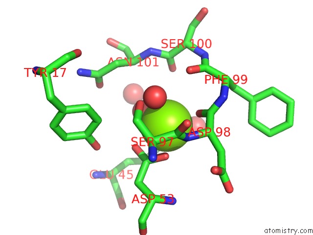
Mono view
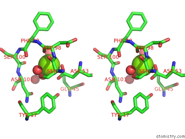
Stereo pair view
Mono view
Stereo pair view
A full contact list of Magnesium with other atoms in the Mg binding
site number 2 of Crystal Structure of the CUB1-Egf Interaction Domain of Complement Protease C1S within 5.0Å range:
|
Reference:
L.A.Gregory,
N.M.Thielens,
G.J.Arlaud,
J.C.Fontecilla-Camps,
C.Gaboriaud.
X-Ray Structure of the CA2+-Binding Interaction Domain of C1S. Insights Into the Assembly of the C1 Complex of Complement J.Biol.Chem. V. 278 32157 2003.
ISSN: ISSN 0021-9258
PubMed: 12788922
DOI: 10.1074/JBC.M305175200
Page generated: Tue Aug 13 10:28:12 2024
ISSN: ISSN 0021-9258
PubMed: 12788922
DOI: 10.1074/JBC.M305175200
Last articles
Fe in 2YXOFe in 2YRS
Fe in 2YXC
Fe in 2YNM
Fe in 2YVJ
Fe in 2YP1
Fe in 2YU2
Fe in 2YU1
Fe in 2YQB
Fe in 2YOO