Magnesium »
PDB 1nzz-1oev »
1oad »
Magnesium in PDB 1oad: Glucose Isomerase From Streptomyces Rubiginosus in P21212 Crystal Form
Enzymatic activity of Glucose Isomerase From Streptomyces Rubiginosus in P21212 Crystal Form
All present enzymatic activity of Glucose Isomerase From Streptomyces Rubiginosus in P21212 Crystal Form:
5.3.1.5;
5.3.1.5;
Protein crystallography data
The structure of Glucose Isomerase From Streptomyces Rubiginosus in P21212 Crystal Form, PDB code: 1oad
was solved by
U.A.Ramagopal,
M.Dauter,
Z.Dauter,
with X-Ray Crystallography technique. A brief refinement statistics is given in the table below:
| Resolution Low / High (Å) | 20.0 / 1.5 |
| Space group | P 21 21 2 |
| Cell size a, b, c (Å), α, β, γ (°) | 98.450, 129.590, 78.330, 90.00, 90.00, 90.00 |
| R / Rfree (%) | 16.3 / 18.6 |
Other elements in 1oad:
The structure of Glucose Isomerase From Streptomyces Rubiginosus in P21212 Crystal Form also contains other interesting chemical elements:
| Manganese | (Mn) | 2 atoms |
Magnesium Binding Sites:
The binding sites of Magnesium atom in the Glucose Isomerase From Streptomyces Rubiginosus in P21212 Crystal Form
(pdb code 1oad). This binding sites where shown within
5.0 Angstroms radius around Magnesium atom.
In total 2 binding sites of Magnesium where determined in the Glucose Isomerase From Streptomyces Rubiginosus in P21212 Crystal Form, PDB code: 1oad:
Jump to Magnesium binding site number: 1; 2;
In total 2 binding sites of Magnesium where determined in the Glucose Isomerase From Streptomyces Rubiginosus in P21212 Crystal Form, PDB code: 1oad:
Jump to Magnesium binding site number: 1; 2;
Magnesium binding site 1 out of 2 in 1oad
Go back to
Magnesium binding site 1 out
of 2 in the Glucose Isomerase From Streptomyces Rubiginosus in P21212 Crystal Form
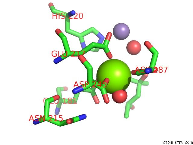
Mono view
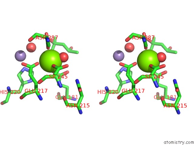
Stereo pair view
Mono view
Stereo pair view
A full contact list of Magnesium with other atoms in the Mg binding
site number 1 of Glucose Isomerase From Streptomyces Rubiginosus in P21212 Crystal Form within 5.0Å range:
|
Magnesium binding site 2 out of 2 in 1oad
Go back to
Magnesium binding site 2 out
of 2 in the Glucose Isomerase From Streptomyces Rubiginosus in P21212 Crystal Form
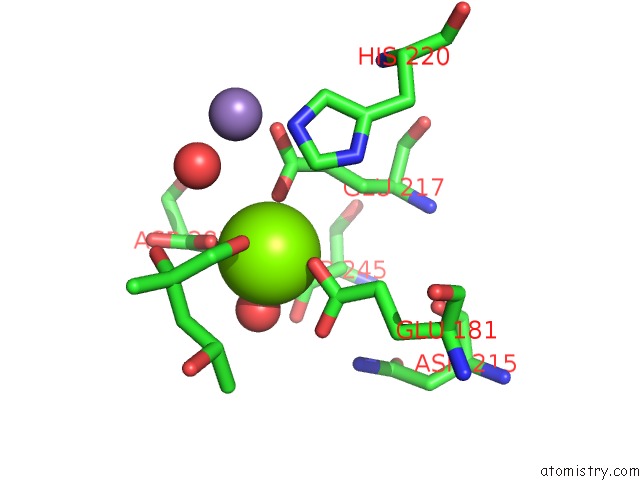
Mono view
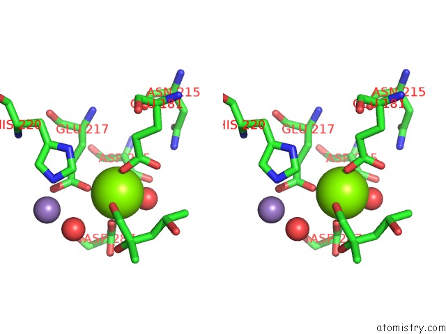
Stereo pair view
Mono view
Stereo pair view
A full contact list of Magnesium with other atoms in the Mg binding
site number 2 of Glucose Isomerase From Streptomyces Rubiginosus in P21212 Crystal Form within 5.0Å range:
|
Reference:
U.A.Ramagopal,
M.Dauter,
Z.Dauter.
Sad Manganese in Two Crystal Forms of Glucose Isomerase Acta Crystallogr.,Sect.D V. 59 868 2003.
ISSN: ISSN 0907-4449
PubMed: 12777803
DOI: 10.1107/S0907444903005663
Page generated: Tue Aug 13 10:35:10 2024
ISSN: ISSN 0907-4449
PubMed: 12777803
DOI: 10.1107/S0907444903005663
Last articles
Zn in 9J0NZn in 9J0O
Zn in 9J0P
Zn in 9FJX
Zn in 9EKB
Zn in 9C0F
Zn in 9CAH
Zn in 9CH0
Zn in 9CH3
Zn in 9CH1