Magnesium »
PDB 1nzz-1oev »
1oe0 »
Magnesium in PDB 1oe0: Crystal Structure of Drosophila Deoxyribonucleoside Kinase in Complex with Dttp
Enzymatic activity of Crystal Structure of Drosophila Deoxyribonucleoside Kinase in Complex with Dttp
All present enzymatic activity of Crystal Structure of Drosophila Deoxyribonucleoside Kinase in Complex with Dttp:
2.7.1.145;
2.7.1.145;
Protein crystallography data
The structure of Crystal Structure of Drosophila Deoxyribonucleoside Kinase in Complex with Dttp, PDB code: 1oe0
was solved by
N.E.Mikkelsen,
K.Johansson,
A.Karlsson,
W.Knecht,
G.Andersen,
J.Piskur,
B.Munch-Petersen,
H.Eklund,
with X-Ray Crystallography technique. A brief refinement statistics is given in the table below:
| Resolution Low / High (Å) | 24.73 / 2.4 |
| Space group | P 1 21 1 |
| Cell size a, b, c (Å), α, β, γ (°) | 67.078, 119.680, 69.512, 90.00, 92.85, 90.00 |
| R / Rfree (%) | 22 / 24.2 |
Magnesium Binding Sites:
The binding sites of Magnesium atom in the Crystal Structure of Drosophila Deoxyribonucleoside Kinase in Complex with Dttp
(pdb code 1oe0). This binding sites where shown within
5.0 Angstroms radius around Magnesium atom.
In total 4 binding sites of Magnesium where determined in the Crystal Structure of Drosophila Deoxyribonucleoside Kinase in Complex with Dttp, PDB code: 1oe0:
Jump to Magnesium binding site number: 1; 2; 3; 4;
In total 4 binding sites of Magnesium where determined in the Crystal Structure of Drosophila Deoxyribonucleoside Kinase in Complex with Dttp, PDB code: 1oe0:
Jump to Magnesium binding site number: 1; 2; 3; 4;
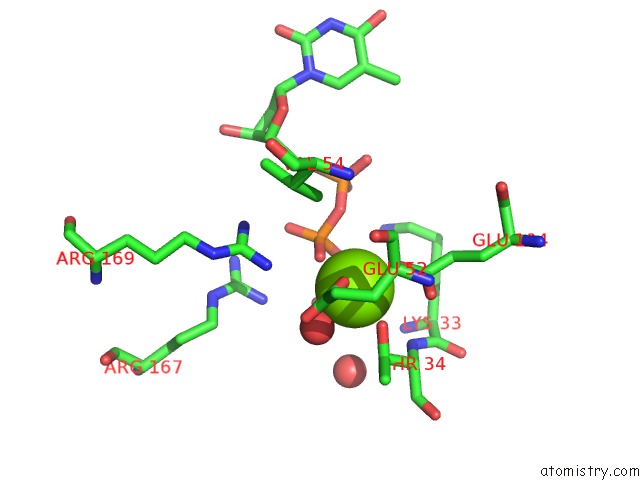
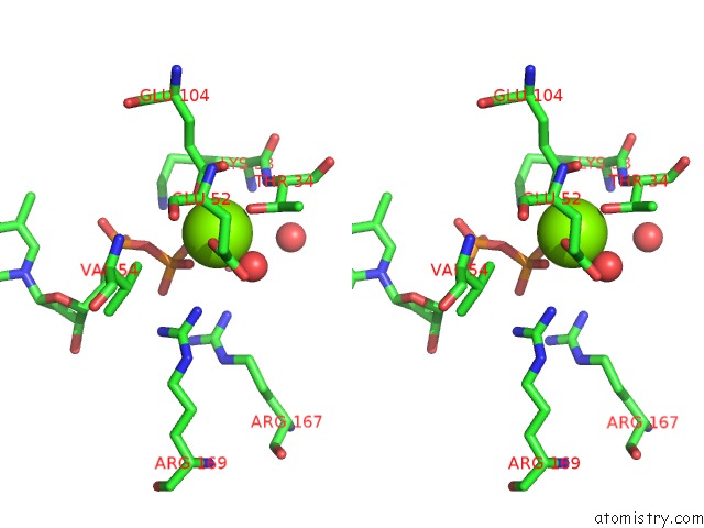
Magnesium binding site 1 out of 4 in 1oe0
Go back to
Magnesium binding site 1 out
of 4 in the Crystal Structure of Drosophila Deoxyribonucleoside Kinase in Complex with Dttp
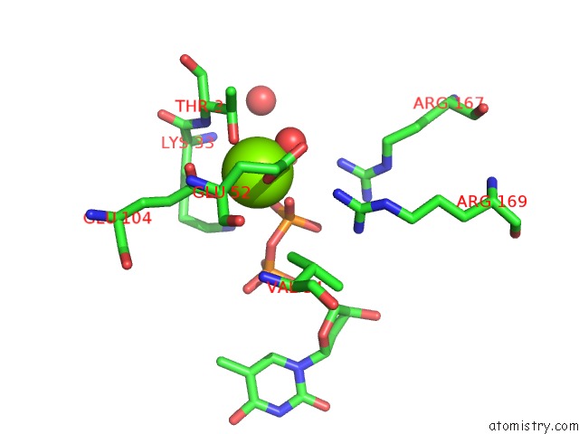
Mono view
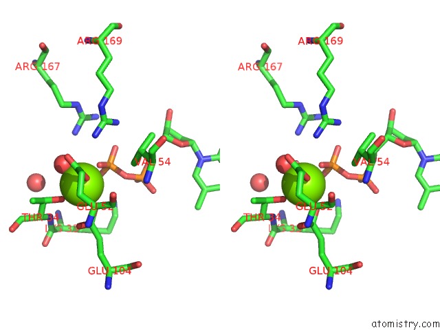
Stereo pair view
Mono view
Stereo pair view
A full contact list of Magnesium with other atoms in the Mg binding
site number 1 of Crystal Structure of Drosophila Deoxyribonucleoside Kinase in Complex with Dttp within 5.0Å range:
|
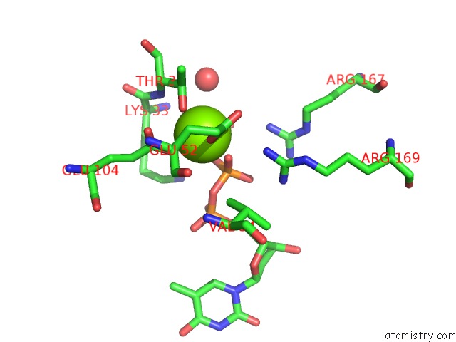
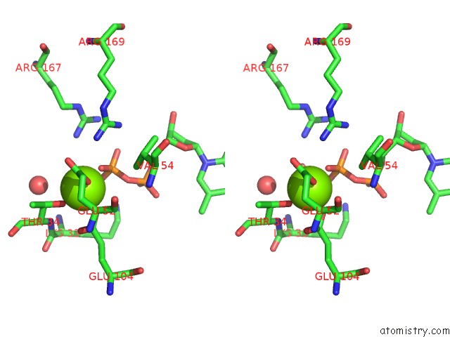
Magnesium binding site 2 out of 4 in 1oe0
Go back to
Magnesium binding site 2 out
of 4 in the Crystal Structure of Drosophila Deoxyribonucleoside Kinase in Complex with Dttp
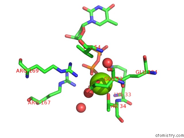
Mono view
Stereo pair view
Mono view
Stereo pair view
A full contact list of Magnesium with other atoms in the Mg binding
site number 2 of Crystal Structure of Drosophila Deoxyribonucleoside Kinase in Complex with Dttp within 5.0Å range:
|
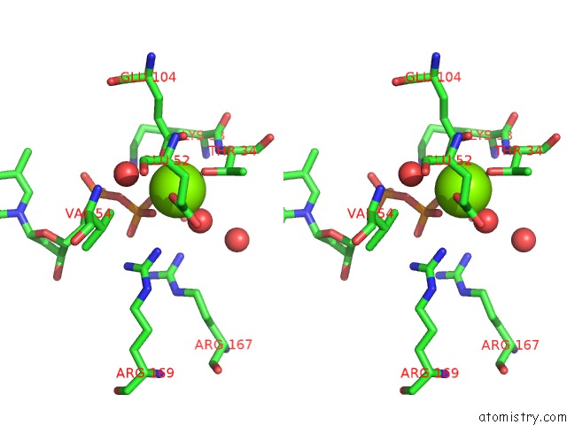
Magnesium binding site 3 out of 4 in 1oe0
Go back to
Magnesium binding site 3 out
of 4 in the Crystal Structure of Drosophila Deoxyribonucleoside Kinase in Complex with Dttp
Mono view
Stereo pair view
Mono view
Stereo pair view
A full contact list of Magnesium with other atoms in the Mg binding
site number 3 of Crystal Structure of Drosophila Deoxyribonucleoside Kinase in Complex with Dttp within 5.0Å range:
|
Magnesium binding site 4 out of 4 in 1oe0
Go back to
Magnesium binding site 4 out
of 4 in the Crystal Structure of Drosophila Deoxyribonucleoside Kinase in Complex with Dttp
Mono view
Stereo pair view
Mono view
Stereo pair view
A full contact list of Magnesium with other atoms in the Mg binding
site number 4 of Crystal Structure of Drosophila Deoxyribonucleoside Kinase in Complex with Dttp within 5.0Å range:
|
Reference:
N.E.Mikkelsen,
K.Johansson,
A.Karlsson,
W.Knecht,
G.Andersen,
J.Piskur,
B.Munch-Petersen,
H.Eklund.
Structural Basis For Feedback Inhibition of the Deoxyribonucleoside Salvage Pathway:Studies of the Drosophila Deoxyribonucleoside Kinase Biochemistry V. 42 5706 2003.
ISSN: ISSN 0006-2960
PubMed: 12741827
DOI: 10.1021/BI0340043
Page generated: Tue Aug 13 10:37:30 2024
ISSN: ISSN 0006-2960
PubMed: 12741827
DOI: 10.1021/BI0340043
Last articles
Zn in 9J0NZn in 9J0O
Zn in 9J0P
Zn in 9FJX
Zn in 9EKB
Zn in 9C0F
Zn in 9CAH
Zn in 9CH0
Zn in 9CH3
Zn in 9CH1