Magnesium »
PDB 1nzz-1oev »
1oeu »
Magnesium in PDB 1oeu: Oxidation State of Protein Tyrosine Phosphatase 1B
Enzymatic activity of Oxidation State of Protein Tyrosine Phosphatase 1B
All present enzymatic activity of Oxidation State of Protein Tyrosine Phosphatase 1B:
3.1.3.48;
3.1.3.48;
Protein crystallography data
The structure of Oxidation State of Protein Tyrosine Phosphatase 1B, PDB code: 1oeu
was solved by
R.L.M.Van Montfort,
M.Congreve,
D.Tisi,
R.Carr,
H.Jhoti,
with X-Ray Crystallography technique. A brief refinement statistics is given in the table below:
| Resolution Low / High (Å) | 76.70 / 2.50 |
| Space group | P 31 2 1 |
| Cell size a, b, c (Å), α, β, γ (°) | 88.914, 88.914, 104.645, 90.00, 90.00, 120.00 |
| R / Rfree (%) | 19.6 / 27 |
Magnesium Binding Sites:
The binding sites of Magnesium atom in the Oxidation State of Protein Tyrosine Phosphatase 1B
(pdb code 1oeu). This binding sites where shown within
5.0 Angstroms radius around Magnesium atom.
In total only one binding site of Magnesium was determined in the Oxidation State of Protein Tyrosine Phosphatase 1B, PDB code: 1oeu:
In total only one binding site of Magnesium was determined in the Oxidation State of Protein Tyrosine Phosphatase 1B, PDB code: 1oeu:
Magnesium binding site 1 out of 1 in 1oeu
Go back to
Magnesium binding site 1 out
of 1 in the Oxidation State of Protein Tyrosine Phosphatase 1B
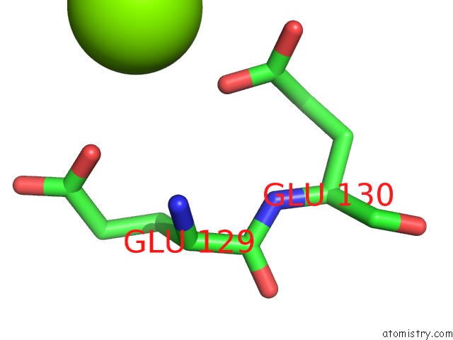
Mono view
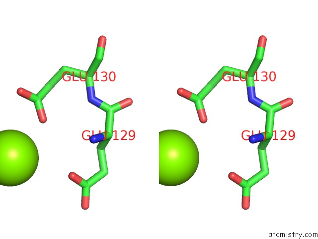
Stereo pair view
Mono view
Stereo pair view
A full contact list of Magnesium with other atoms in the Mg binding
site number 1 of Oxidation State of Protein Tyrosine Phosphatase 1B within 5.0Å range:
|
Reference:
R.L.Van Montfort,
M.Congreve,
D.Tisi,
R.Carr,
H.Jhoti.
Oxidation State of the Active-Site Cysteine in Protein Tyrosine Phosphatase 1B. Nature V. 423 773 2003.
ISSN: ISSN 0028-0836
PubMed: 12802339
DOI: 10.1038/NATURE01681
Page generated: Tue Aug 13 10:37:41 2024
ISSN: ISSN 0028-0836
PubMed: 12802339
DOI: 10.1038/NATURE01681
Last articles
Zn in 9J0NZn in 9J0O
Zn in 9J0P
Zn in 9FJX
Zn in 9EKB
Zn in 9C0F
Zn in 9CAH
Zn in 9CH0
Zn in 9CH3
Zn in 9CH1