Magnesium »
PDB 1ofh-1oyj »
1ol5 »
Magnesium in PDB 1ol5: Structure of Aurora-A 122-403, Phosphorylated on THR287, THR288 and Bound to TPX2 1-43
Enzymatic activity of Structure of Aurora-A 122-403, Phosphorylated on THR287, THR288 and Bound to TPX2 1-43
All present enzymatic activity of Structure of Aurora-A 122-403, Phosphorylated on THR287, THR288 and Bound to TPX2 1-43:
2.7.1.37;
2.7.1.37;
Protein crystallography data
The structure of Structure of Aurora-A 122-403, Phosphorylated on THR287, THR288 and Bound to TPX2 1-43, PDB code: 1ol5
was solved by
R.Bayliss,
E.Conti,
with X-Ray Crystallography technique. A brief refinement statistics is given in the table below:
| Resolution Low / High (Å) | 40 / 2.5 |
| Space group | P 21 21 21 |
| Cell size a, b, c (Å), α, β, γ (°) | 59.630, 81.720, 83.050, 90.00, 90.00, 90.00 |
| R / Rfree (%) | 19.4 / 25.2 |
Magnesium Binding Sites:
The binding sites of Magnesium atom in the Structure of Aurora-A 122-403, Phosphorylated on THR287, THR288 and Bound to TPX2 1-43
(pdb code 1ol5). This binding sites where shown within
5.0 Angstroms radius around Magnesium atom.
In total 3 binding sites of Magnesium where determined in the Structure of Aurora-A 122-403, Phosphorylated on THR287, THR288 and Bound to TPX2 1-43, PDB code: 1ol5:
Jump to Magnesium binding site number: 1; 2; 3;
In total 3 binding sites of Magnesium where determined in the Structure of Aurora-A 122-403, Phosphorylated on THR287, THR288 and Bound to TPX2 1-43, PDB code: 1ol5:
Jump to Magnesium binding site number: 1; 2; 3;
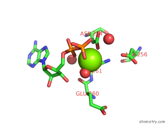
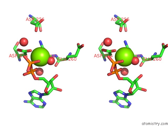
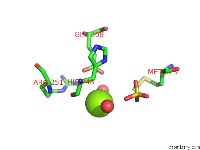
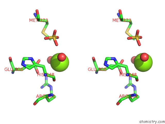
Magnesium binding site 1 out of 3 in 1ol5
Go back to
Magnesium binding site 1 out
of 3 in the Structure of Aurora-A 122-403, Phosphorylated on THR287, THR288 and Bound to TPX2 1-43
Mono view
Stereo pair view
Mono view
Stereo pair view
A full contact list of Magnesium with other atoms in the Mg binding
site number 1 of Structure of Aurora-A 122-403, Phosphorylated on THR287, THR288 and Bound to TPX2 1-43 within 5.0Å range:
|
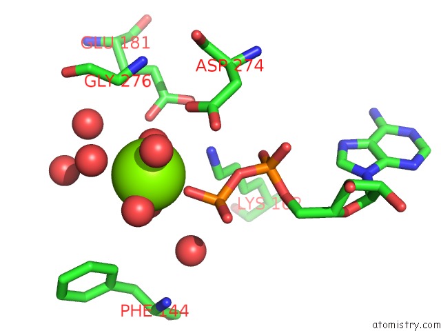
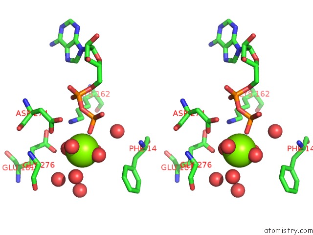
Magnesium binding site 2 out of 3 in 1ol5
Go back to
Magnesium binding site 2 out
of 3 in the Structure of Aurora-A 122-403, Phosphorylated on THR287, THR288 and Bound to TPX2 1-43
Mono view
Stereo pair view
Mono view
Stereo pair view
A full contact list of Magnesium with other atoms in the Mg binding
site number 2 of Structure of Aurora-A 122-403, Phosphorylated on THR287, THR288 and Bound to TPX2 1-43 within 5.0Å range:
|
Magnesium binding site 3 out of 3 in 1ol5
Go back to
Magnesium binding site 3 out
of 3 in the Structure of Aurora-A 122-403, Phosphorylated on THR287, THR288 and Bound to TPX2 1-43
Mono view
Stereo pair view
Mono view
Stereo pair view
A full contact list of Magnesium with other atoms in the Mg binding
site number 3 of Structure of Aurora-A 122-403, Phosphorylated on THR287, THR288 and Bound to TPX2 1-43 within 5.0Å range:
|
Reference:
R.Bayliss,
T.Sardon,
I.Vernos,
E.Conti.
Structural Basis of Aurora-A Activation By TPX2 at the Mitotic Spindle Mol.Cell V. 12 851 2003.
ISSN: ISSN 1097-2765
PubMed: 14580337
DOI: 10.1016/S1097-2765(03)00392-7
Page generated: Tue Aug 13 10:40:32 2024
ISSN: ISSN 1097-2765
PubMed: 14580337
DOI: 10.1016/S1097-2765(03)00392-7
Last articles
Zn in 9J0NZn in 9J0O
Zn in 9J0P
Zn in 9FJX
Zn in 9EKB
Zn in 9C0F
Zn in 9CAH
Zn in 9CH0
Zn in 9CH3
Zn in 9CH1