Magnesium »
PDB 1ofh-1oyj »
1onw »
Magnesium in PDB 1onw: Crystal Structure of Isoaspartyl Dipeptidase From E. Coli
Protein crystallography data
The structure of Crystal Structure of Isoaspartyl Dipeptidase From E. Coli, PDB code: 1onw
was solved by
J.B.Thoden,
R.Marti-Arbona,
F.M.Raushel,
H.M.Holden,
with X-Ray Crystallography technique. A brief refinement statistics is given in the table below:
| Resolution Low / High (Å) | 30.00 / 1.65 |
| Space group | P 4 21 2 |
| Cell size a, b, c (Å), α, β, γ (°) | 116.700, 116.700, 138.500, 90.00, 90.00, 90.00 |
| R / Rfree (%) | n/a / n/a |
Other elements in 1onw:
The structure of Crystal Structure of Isoaspartyl Dipeptidase From E. Coli also contains other interesting chemical elements:
| Zinc | (Zn) | 4 atoms |
| Chlorine | (Cl) | 3 atoms |
| Sodium | (Na) | 1 atom |
Magnesium Binding Sites:
The binding sites of Magnesium atom in the Crystal Structure of Isoaspartyl Dipeptidase From E. Coli
(pdb code 1onw). This binding sites where shown within
5.0 Angstroms radius around Magnesium atom.
In total only one binding site of Magnesium was determined in the Crystal Structure of Isoaspartyl Dipeptidase From E. Coli, PDB code: 1onw:
In total only one binding site of Magnesium was determined in the Crystal Structure of Isoaspartyl Dipeptidase From E. Coli, PDB code: 1onw:
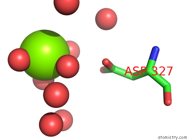
Magnesium binding site 1 out of 1 in 1onw
Go back to
Magnesium binding site 1 out
of 1 in the Crystal Structure of Isoaspartyl Dipeptidase From E. Coli
Mono view

Stereo pair view
Mono view
Stereo pair view
A full contact list of Magnesium with other atoms in the Mg binding
site number 1 of Crystal Structure of Isoaspartyl Dipeptidase From E. Coli within 5.0Å range:
|
Reference:
J.B.Thoden,
R.Marti-Arbona,
F.M.Raushel,
H.M.Holden.
High Resolution X-Ray Structure of Isoaspartyl Dipeptidase From Escherichia Coli Biochemistry V. 42 4874 2003.
ISSN: ISSN 0006-2960
PubMed: 12718528
DOI: 10.1021/BI034233P
Page generated: Tue Aug 13 10:41:44 2024
ISSN: ISSN 0006-2960
PubMed: 12718528
DOI: 10.1021/BI034233P
Last articles
Zn in 9J0NZn in 9J0O
Zn in 9J0P
Zn in 9FJX
Zn in 9EKB
Zn in 9C0F
Zn in 9CAH
Zn in 9CH0
Zn in 9CH3
Zn in 9CH1