Magnesium »
PDB 1ozf-1php »
1p62 »
Magnesium in PDB 1p62: Structure of Human Dck Complexed with Gemcitabine and Adp-Mg
Enzymatic activity of Structure of Human Dck Complexed with Gemcitabine and Adp-Mg
All present enzymatic activity of Structure of Human Dck Complexed with Gemcitabine and Adp-Mg:
2.7.1.74;
2.7.1.74;
Protein crystallography data
The structure of Structure of Human Dck Complexed with Gemcitabine and Adp-Mg, PDB code: 1p62
was solved by
E.Sabini,
S.Ort,
C.Monnerjahn,
M.Konrad,
A.Lavie,
with X-Ray Crystallography technique. A brief refinement statistics is given in the table below:
| Resolution Low / High (Å) | 20.00 / 1.90 |
| Space group | P 43 21 2 |
| Cell size a, b, c (Å), α, β, γ (°) | 81.200, 81.200, 94.600, 90.00, 90.00, 90.00 |
| R / Rfree (%) | 17.9 / 20.7 |
Other elements in 1p62:
The structure of Structure of Human Dck Complexed with Gemcitabine and Adp-Mg also contains other interesting chemical elements:
| Fluorine | (F) | 2 atoms |
Magnesium Binding Sites:
The binding sites of Magnesium atom in the Structure of Human Dck Complexed with Gemcitabine and Adp-Mg
(pdb code 1p62). This binding sites where shown within
5.0 Angstroms radius around Magnesium atom.
In total only one binding site of Magnesium was determined in the Structure of Human Dck Complexed with Gemcitabine and Adp-Mg, PDB code: 1p62:
In total only one binding site of Magnesium was determined in the Structure of Human Dck Complexed with Gemcitabine and Adp-Mg, PDB code: 1p62:
Magnesium binding site 1 out of 1 in 1p62
Go back to
Magnesium binding site 1 out
of 1 in the Structure of Human Dck Complexed with Gemcitabine and Adp-Mg
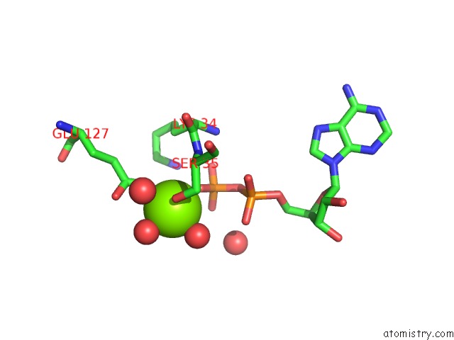
Mono view
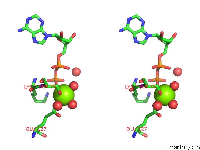
Stereo pair view
Mono view
Stereo pair view
A full contact list of Magnesium with other atoms in the Mg binding
site number 1 of Structure of Human Dck Complexed with Gemcitabine and Adp-Mg within 5.0Å range:
|
Reference:
E.Sabini,
S.Ort,
C.Monnerjahn,
M.Konrad,
A.Lavie.
Structure of Human Dck Suggests Strategies to Improve Anticancer and Antiviral Therapy Nat.Struct.Biol. V. 10 513 2003.
ISSN: ISSN 1072-8368
PubMed: 12808445
DOI: 10.1038/NSB942
Page generated: Tue Aug 13 10:49:28 2024
ISSN: ISSN 1072-8368
PubMed: 12808445
DOI: 10.1038/NSB942
Last articles
Zn in 9J0NZn in 9J0O
Zn in 9J0P
Zn in 9FJX
Zn in 9EKB
Zn in 9C0F
Zn in 9CAH
Zn in 9CH0
Zn in 9CH3
Zn in 9CH1