Magnesium »
PDB 1pi3-1q24 »
1pst »
Magnesium in PDB 1pst: Crystallographic Analyses of Site-Directed Mutants of the Photosynthetic Reaction Center From Rhodobacter Sphaeroides
Protein crystallography data
The structure of Crystallographic Analyses of Site-Directed Mutants of the Photosynthetic Reaction Center From Rhodobacter Sphaeroides, PDB code: 1pst
was solved by
A.J.Chirino,
G.Feher,
D.C.Rees,
with X-Ray Crystallography technique. A brief refinement statistics is given in the table below:
| Resolution Low / High (Å) | N/A / 3.00 |
| Space group | P 21 21 21 |
| Cell size a, b, c (Å), α, β, γ (°) | 138.000, 77.500, 141.800, 90.00, 90.00, 90.00 |
| R / Rfree (%) | 21.8 / n/a |
Other elements in 1pst:
The structure of Crystallographic Analyses of Site-Directed Mutants of the Photosynthetic Reaction Center From Rhodobacter Sphaeroides also contains other interesting chemical elements:
| Iron | (Fe) | 1 atom |
Magnesium Binding Sites:
The binding sites of Magnesium atom in the Crystallographic Analyses of Site-Directed Mutants of the Photosynthetic Reaction Center From Rhodobacter Sphaeroides
(pdb code 1pst). This binding sites where shown within
5.0 Angstroms radius around Magnesium atom.
In total 3 binding sites of Magnesium where determined in the Crystallographic Analyses of Site-Directed Mutants of the Photosynthetic Reaction Center From Rhodobacter Sphaeroides, PDB code: 1pst:
Jump to Magnesium binding site number: 1; 2; 3;
In total 3 binding sites of Magnesium where determined in the Crystallographic Analyses of Site-Directed Mutants of the Photosynthetic Reaction Center From Rhodobacter Sphaeroides, PDB code: 1pst:
Jump to Magnesium binding site number: 1; 2; 3;
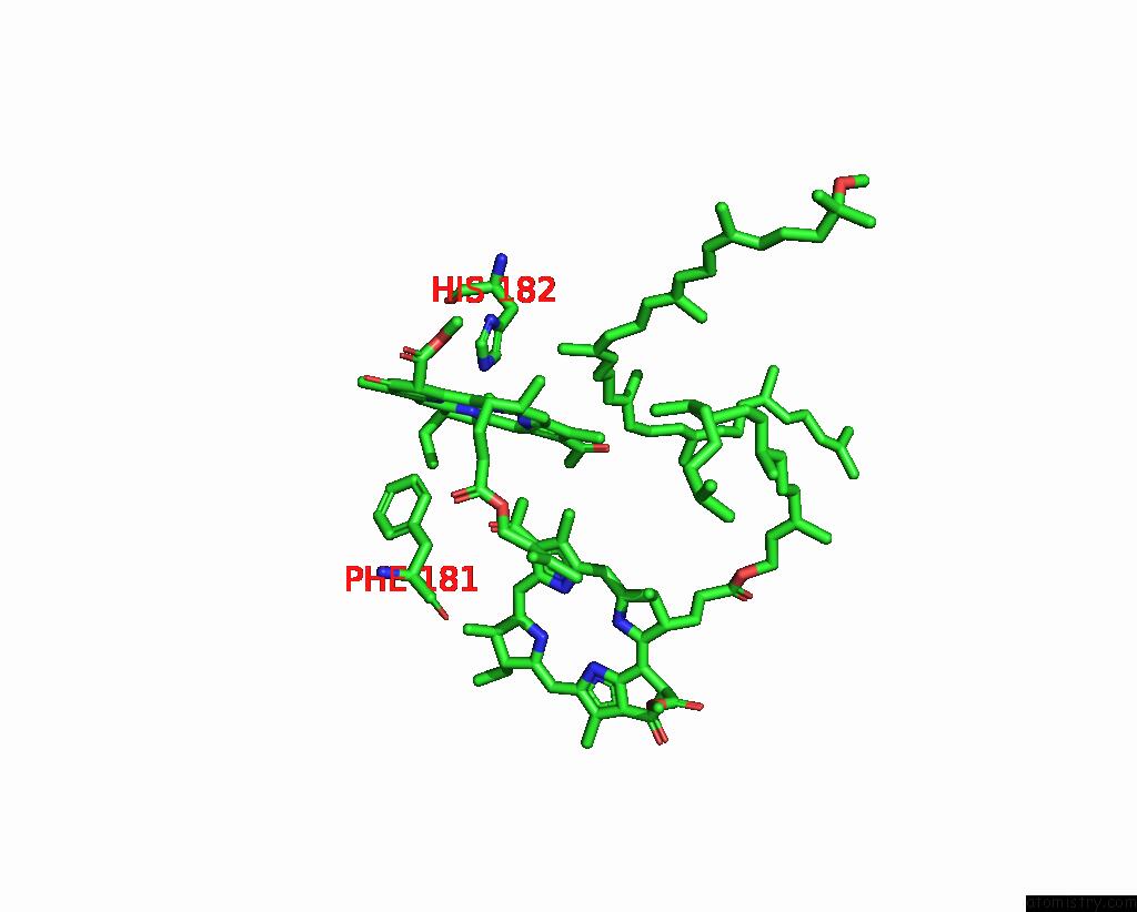
Magnesium binding site 1 out of 3 in 1pst
Go back to
Magnesium binding site 1 out
of 3 in the Crystallographic Analyses of Site-Directed Mutants of the Photosynthetic Reaction Center From Rhodobacter Sphaeroides
Mono view

Stereo pair view
Mono view
Stereo pair view
A full contact list of Magnesium with other atoms in the Mg binding
site number 1 of Crystallographic Analyses of Site-Directed Mutants of the Photosynthetic Reaction Center From Rhodobacter Sphaeroides within 5.0Å range:
|
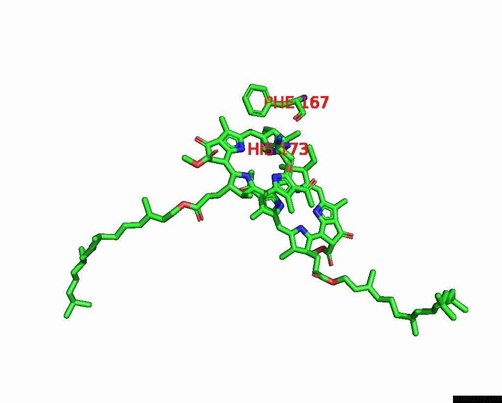
Magnesium binding site 2 out of 3 in 1pst
Go back to
Magnesium binding site 2 out
of 3 in the Crystallographic Analyses of Site-Directed Mutants of the Photosynthetic Reaction Center From Rhodobacter Sphaeroides
Mono view
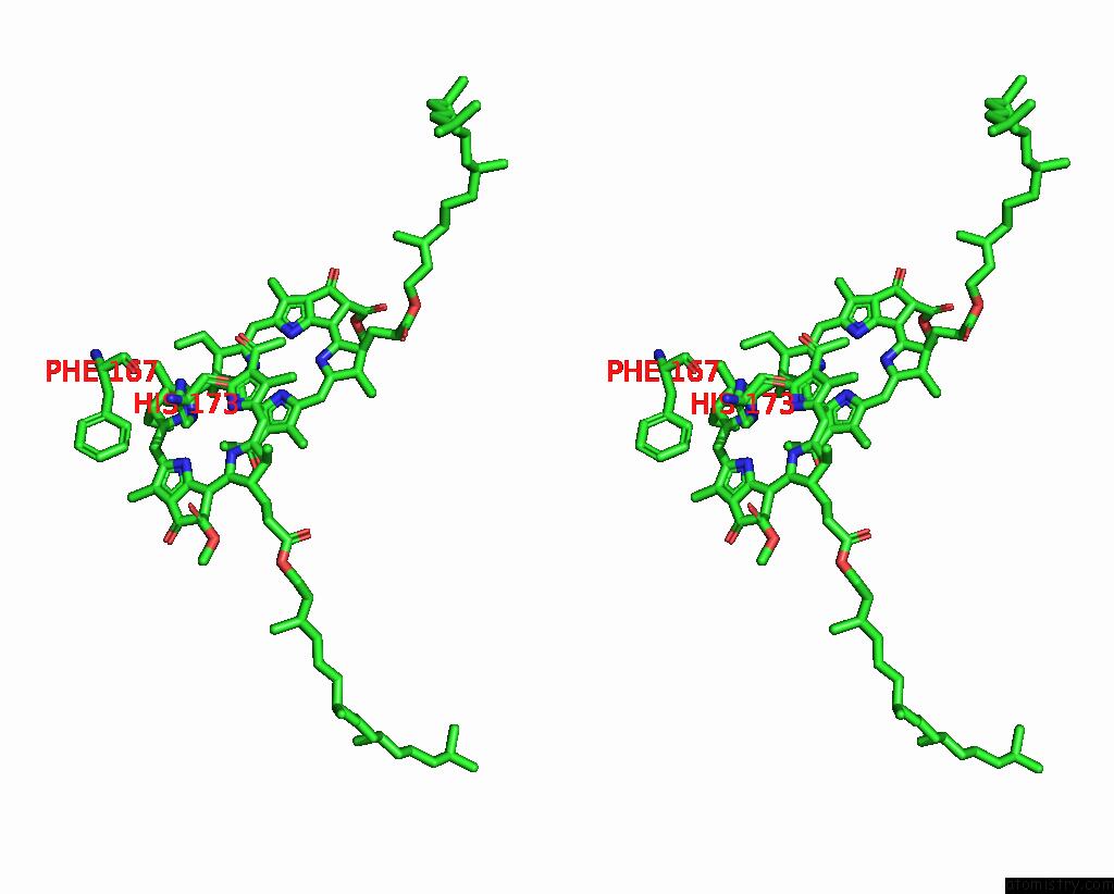
Stereo pair view
Mono view
Stereo pair view
A full contact list of Magnesium with other atoms in the Mg binding
site number 2 of Crystallographic Analyses of Site-Directed Mutants of the Photosynthetic Reaction Center From Rhodobacter Sphaeroides within 5.0Å range:
|
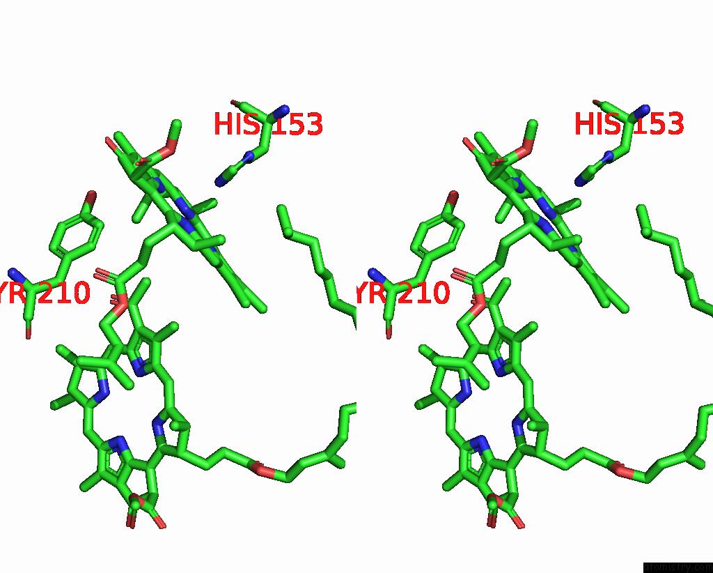
Magnesium binding site 3 out of 3 in 1pst
Go back to
Magnesium binding site 3 out
of 3 in the Crystallographic Analyses of Site-Directed Mutants of the Photosynthetic Reaction Center From Rhodobacter Sphaeroides

Mono view
Stereo pair view
Mono view
Stereo pair view
A full contact list of Magnesium with other atoms in the Mg binding
site number 3 of Crystallographic Analyses of Site-Directed Mutants of the Photosynthetic Reaction Center From Rhodobacter Sphaeroides within 5.0Å range:
|
Reference:
A.J.Chirino,
E.J.Lous,
M.Huber,
J.P.Allen,
C.C.Schenck,
M.L.Paddock,
G.Feher,
D.C.Rees.
Crystallographic Analyses of Site-Directed Mutants of the Photosynthetic Reaction Center From Rhodobacter Sphaeroides. Biochemistry V. 33 4584 1994.
ISSN: ISSN 0006-2960
PubMed: 8161514
DOI: 10.1021/BI00181A020
Page generated: Tue Aug 13 10:55:17 2024
ISSN: ISSN 0006-2960
PubMed: 8161514
DOI: 10.1021/BI00181A020
Last articles
Zn in 9MJ5Zn in 9HNW
Zn in 9G0L
Zn in 9FNE
Zn in 9DZN
Zn in 9E0I
Zn in 9D32
Zn in 9DAK
Zn in 8ZXC
Zn in 8ZUF