Magnesium »
PDB 1q3b-1qc1 »
1q91 »
Magnesium in PDB 1q91: Crystal Structure of Human Mitochondrial Deoxyribonucleotidase in Complex with the Inhibitor Dpb-T
Enzymatic activity of Crystal Structure of Human Mitochondrial Deoxyribonucleotidase in Complex with the Inhibitor Dpb-T
All present enzymatic activity of Crystal Structure of Human Mitochondrial Deoxyribonucleotidase in Complex with the Inhibitor Dpb-T:
3.1.3.5;
3.1.3.5;
Protein crystallography data
The structure of Crystal Structure of Human Mitochondrial Deoxyribonucleotidase in Complex with the Inhibitor Dpb-T, PDB code: 1q91
was solved by
A.Rinaldo-Matthis,
C.Rampazzo,
J.Balzarini,
P.Reichard,
V.Bianchi,
P.Nordlund,
with X-Ray Crystallography technique. A brief refinement statistics is given in the table below:
| Resolution Low / High (Å) | 19.77 / 1.60 |
| Space group | P 43 21 2 |
| Cell size a, b, c (Å), α, β, γ (°) | 73.651, 73.651, 106.830, 90.00, 90.00, 90.00 |
| R / Rfree (%) | 18.6 / 20.6 |
Magnesium Binding Sites:
The binding sites of Magnesium atom in the Crystal Structure of Human Mitochondrial Deoxyribonucleotidase in Complex with the Inhibitor Dpb-T
(pdb code 1q91). This binding sites where shown within
5.0 Angstroms radius around Magnesium atom.
In total only one binding site of Magnesium was determined in the Crystal Structure of Human Mitochondrial Deoxyribonucleotidase in Complex with the Inhibitor Dpb-T, PDB code: 1q91:
In total only one binding site of Magnesium was determined in the Crystal Structure of Human Mitochondrial Deoxyribonucleotidase in Complex with the Inhibitor Dpb-T, PDB code: 1q91:
Magnesium binding site 1 out of 1 in 1q91
Go back to
Magnesium binding site 1 out
of 1 in the Crystal Structure of Human Mitochondrial Deoxyribonucleotidase in Complex with the Inhibitor Dpb-T
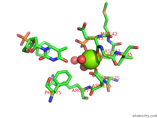
Mono view
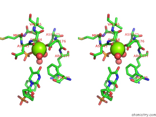
Stereo pair view
Mono view
Stereo pair view
A full contact list of Magnesium with other atoms in the Mg binding
site number 1 of Crystal Structure of Human Mitochondrial Deoxyribonucleotidase in Complex with the Inhibitor Dpb-T within 5.0Å range:
|
Reference:
A.Rinaldo-Matthis,
C.Rampazzo,
J.Balzarini,
P.Reichard,
V.Bianchi,
P.Nordlund.
Crystal Structures of the Mitochondrial Deoxyribonucleotidase in Complex with Two Specific Inhibitors Mol.Pharmacol. V. 65 860 2004.
ISSN: ISSN 0026-895X
PubMed: 15044615
DOI: 10.1124/MOL.65.4.860
Page generated: Sun Aug 10 02:58:49 2025
ISSN: ISSN 0026-895X
PubMed: 15044615
DOI: 10.1124/MOL.65.4.860
Last articles
Mg in 4LF2Mg in 4LF1
Mg in 4LEM
Mg in 4LCK
Mg in 4LE0
Mg in 4LDZ
Mg in 4LDT
Mg in 4LA7
Mg in 4LDJ
Mg in 4LC8