Magnesium »
PDB 1q3b-1qc1 »
1qc1 »
Magnesium in PDB 1qc1: Crystal Structure of the Self-Fitted B-Dna Decamer D(Ccgccggcgg)
Protein crystallography data
The structure of Crystal Structure of the Self-Fitted B-Dna Decamer D(Ccgccggcgg), PDB code: 1qc1
was solved by
Y.Timsit,
D.Moras,
with X-Ray Crystallography technique. A brief refinement statistics is given in the table below:
| Resolution Low / High (Å) | 7.00 / 2.50 |
| Space group | H 3 |
| Cell size a, b, c (Å), α, β, γ (°) | 53.700, 53.700, 45.000, 90.00, 90.00, 120.00 |
| R / Rfree (%) | n/a / n/a |
Magnesium Binding Sites:
The binding sites of Magnesium atom in the Crystal Structure of the Self-Fitted B-Dna Decamer D(Ccgccggcgg)
(pdb code 1qc1). This binding sites where shown within
5.0 Angstroms radius around Magnesium atom.
In total 4 binding sites of Magnesium where determined in the Crystal Structure of the Self-Fitted B-Dna Decamer D(Ccgccggcgg), PDB code: 1qc1:
Jump to Magnesium binding site number: 1; 2; 3; 4;
In total 4 binding sites of Magnesium where determined in the Crystal Structure of the Self-Fitted B-Dna Decamer D(Ccgccggcgg), PDB code: 1qc1:
Jump to Magnesium binding site number: 1; 2; 3; 4;
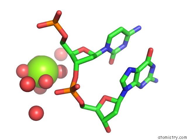
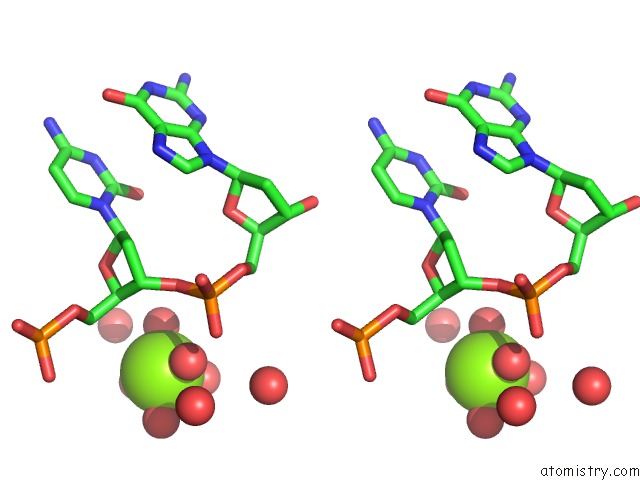
Magnesium binding site 1 out of 4 in 1qc1
Go back to
Magnesium binding site 1 out
of 4 in the Crystal Structure of the Self-Fitted B-Dna Decamer D(Ccgccggcgg)
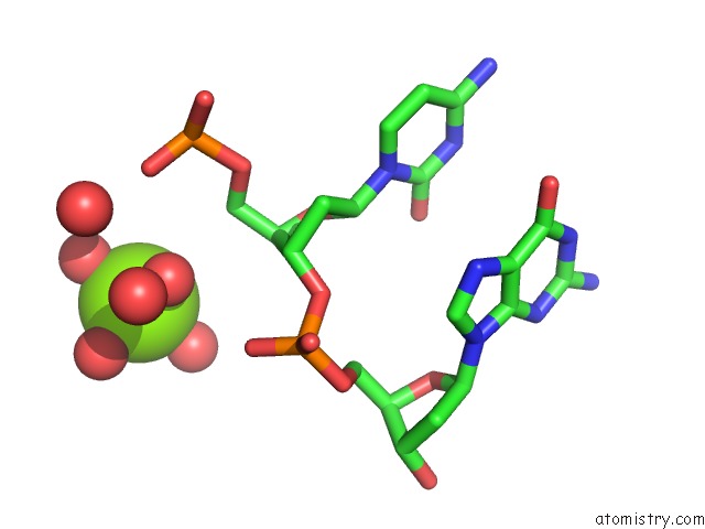
Mono view
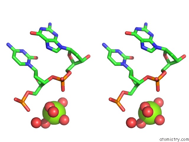
Stereo pair view
Mono view
Stereo pair view
A full contact list of Magnesium with other atoms in the Mg binding
site number 1 of Crystal Structure of the Self-Fitted B-Dna Decamer D(Ccgccggcgg) within 5.0Å range:
|
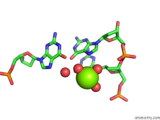
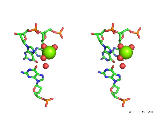
Magnesium binding site 2 out of 4 in 1qc1
Go back to
Magnesium binding site 2 out
of 4 in the Crystal Structure of the Self-Fitted B-Dna Decamer D(Ccgccggcgg)
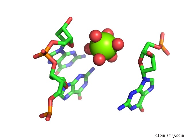
Mono view
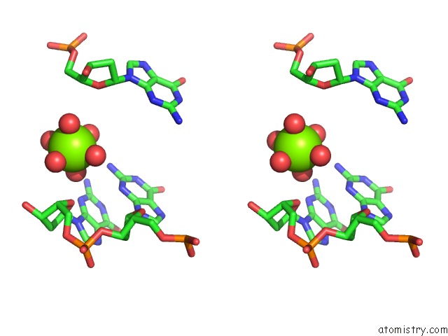
Stereo pair view
Mono view
Stereo pair view
A full contact list of Magnesium with other atoms in the Mg binding
site number 2 of Crystal Structure of the Self-Fitted B-Dna Decamer D(Ccgccggcgg) within 5.0Å range:
|
Magnesium binding site 3 out of 4 in 1qc1
Go back to
Magnesium binding site 3 out
of 4 in the Crystal Structure of the Self-Fitted B-Dna Decamer D(Ccgccggcgg)
Mono view
Stereo pair view
Mono view
Stereo pair view
A full contact list of Magnesium with other atoms in the Mg binding
site number 3 of Crystal Structure of the Self-Fitted B-Dna Decamer D(Ccgccggcgg) within 5.0Å range:
|
Magnesium binding site 4 out of 4 in 1qc1
Go back to
Magnesium binding site 4 out
of 4 in the Crystal Structure of the Self-Fitted B-Dna Decamer D(Ccgccggcgg)
Mono view
Stereo pair view
Mono view
Stereo pair view
A full contact list of Magnesium with other atoms in the Mg binding
site number 4 of Crystal Structure of the Self-Fitted B-Dna Decamer D(Ccgccggcgg) within 5.0Å range:
|
Reference:
Y.Timsit,
D.Moras.
Dna Self-Fitting: the Double Helix Directs the Geometry of Its Supramolecular Assembly Embo J. V. 13 2737 1994.
ISSN: ISSN 0261-4189
PubMed: 8026458
Page generated: Tue Aug 13 11:16:55 2024
ISSN: ISSN 0261-4189
PubMed: 8026458
Last articles
Zn in 9J0NZn in 9J0O
Zn in 9J0P
Zn in 9FJX
Zn in 9EKB
Zn in 9C0F
Zn in 9CAH
Zn in 9CH0
Zn in 9CH3
Zn in 9CH1