Magnesium »
PDB 1qc5-1qsh »
1qf8 »
Magnesium in PDB 1qf8: Truncated Form of Casein Kinase II Beta Subunit (2-182) From Homo Sapiens
Enzymatic activity of Truncated Form of Casein Kinase II Beta Subunit (2-182) From Homo Sapiens
All present enzymatic activity of Truncated Form of Casein Kinase II Beta Subunit (2-182) From Homo Sapiens:
2.7.1.37;
2.7.1.37;
Protein crystallography data
The structure of Truncated Form of Casein Kinase II Beta Subunit (2-182) From Homo Sapiens, PDB code: 1qf8
was solved by
L.Chantalat,
D.Leroy,
O.Filhol,
A.Nueda,
M.J.Benitez,
E.Chambaz,
C.Cochet,
O.Dideberg,
with X-Ray Crystallography technique. A brief refinement statistics is given in the table below:
| Resolution Low / High (Å) | 20.00 / 1.74 |
| Space group | P 42 21 2 |
| Cell size a, b, c (Å), α, β, γ (°) | 132.230, 132.230, 63.780, 90.00, 90.00, 90.00 |
| R / Rfree (%) | 19.4 / 21.9 |
Other elements in 1qf8:
The structure of Truncated Form of Casein Kinase II Beta Subunit (2-182) From Homo Sapiens also contains other interesting chemical elements:
| Zinc | (Zn) | 2 atoms |
Magnesium Binding Sites:
The binding sites of Magnesium atom in the Truncated Form of Casein Kinase II Beta Subunit (2-182) From Homo Sapiens
(pdb code 1qf8). This binding sites where shown within
5.0 Angstroms radius around Magnesium atom.
In total only one binding site of Magnesium was determined in the Truncated Form of Casein Kinase II Beta Subunit (2-182) From Homo Sapiens, PDB code: 1qf8:
In total only one binding site of Magnesium was determined in the Truncated Form of Casein Kinase II Beta Subunit (2-182) From Homo Sapiens, PDB code: 1qf8:
Magnesium binding site 1 out of 1 in 1qf8
Go back to
Magnesium binding site 1 out
of 1 in the Truncated Form of Casein Kinase II Beta Subunit (2-182) From Homo Sapiens
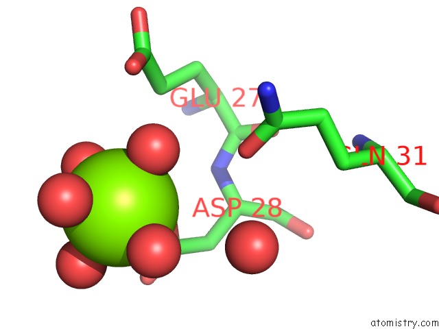
Mono view
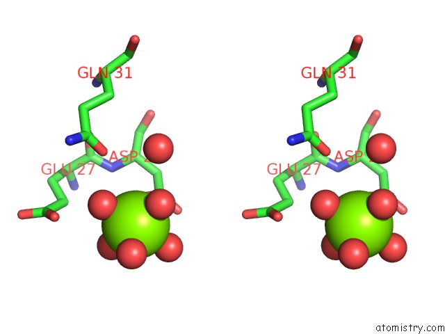
Stereo pair view
Mono view
Stereo pair view
A full contact list of Magnesium with other atoms in the Mg binding
site number 1 of Truncated Form of Casein Kinase II Beta Subunit (2-182) From Homo Sapiens within 5.0Å range:
|
Reference:
L.Chantalat,
D.Leroy,
O.Filhol,
A.Nueda,
M.J.Benitez,
E.M.Chambaz,
C.Cochet,
O.Dideberg.
Crystal Structure of the Human Protein Kinase CK2 Regulatory Subunit Reveals Its Zinc Finger-Mediated Dimerization. Embo J. V. 18 2930 1999.
ISSN: ISSN 0261-4189
PubMed: 10357806
DOI: 10.1093/EMBOJ/18.11.2930
Page generated: Tue Aug 13 11:49:34 2024
ISSN: ISSN 0261-4189
PubMed: 10357806
DOI: 10.1093/EMBOJ/18.11.2930
Last articles
Zn in 9MJ5Zn in 9HNW
Zn in 9G0L
Zn in 9FNE
Zn in 9DZN
Zn in 9E0I
Zn in 9D32
Zn in 9DAK
Zn in 8ZXC
Zn in 8ZUF