Magnesium »
PDB 1qc5-1qsh »
1qhg »
Magnesium in PDB 1qhg: Structure of Dna Helicase Mutant with Adpnp
Protein crystallography data
The structure of Structure of Dna Helicase Mutant with Adpnp, PDB code: 1qhg
was solved by
P.Soultanas,
M.S.Dillingham,
S.S.Velankar,
D.B.Wigley,
with X-Ray Crystallography technique. A brief refinement statistics is given in the table below:
| Resolution Low / High (Å) | 10.00 / 2.50 |
| Space group | P 65 |
| Cell size a, b, c (Å), α, β, γ (°) | 138.098, 138.098, 111.086, 90.00, 90.00, 120.00 |
| R / Rfree (%) | 22 / 26.9 |
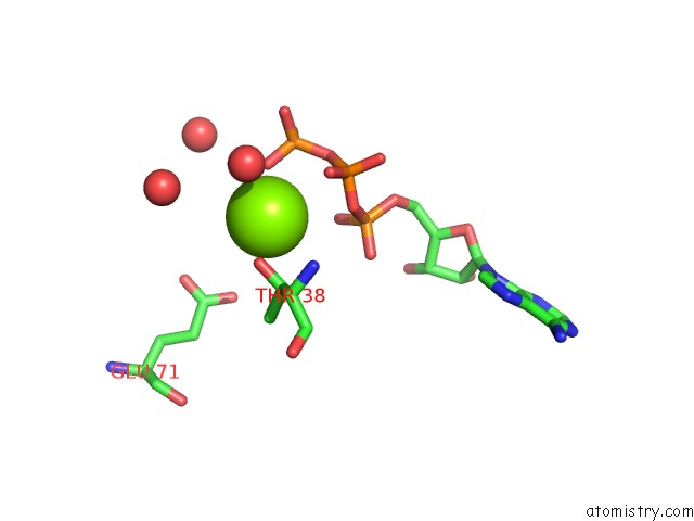
Magnesium Binding Sites:
The binding sites of Magnesium atom in the Structure of Dna Helicase Mutant with Adpnp
(pdb code 1qhg). This binding sites where shown within
5.0 Angstroms radius around Magnesium atom.
In total only one binding site of Magnesium was determined in the Structure of Dna Helicase Mutant with Adpnp, PDB code: 1qhg:
In total only one binding site of Magnesium was determined in the Structure of Dna Helicase Mutant with Adpnp, PDB code: 1qhg:
Magnesium binding site 1 out of 1 in 1qhg
Go back to
Magnesium binding site 1 out
of 1 in the Structure of Dna Helicase Mutant with Adpnp
Mono view

Stereo pair view
Mono view
Stereo pair view
A full contact list of Magnesium with other atoms in the Mg binding
site number 1 of Structure of Dna Helicase Mutant with Adpnp within 5.0Å range:
|
Reference:
P.Soultanas,
M.S.Dillingham,
S.S.Velankar,
D.B.Wigley.
Dna Binding Mediates Conformational Changes and Metal Ion Coordination in the Active Site of Pcra Helicase. J.Mol.Biol. V. 290 137 1999.
ISSN: ISSN 0022-2836
PubMed: 10388562
DOI: 10.1006/JMBI.1999.2873
Page generated: Tue Aug 13 11:51:40 2024
ISSN: ISSN 0022-2836
PubMed: 10388562
DOI: 10.1006/JMBI.1999.2873
Last articles
Ca in 5PB6Ca in 5PB5
Ca in 5PB4
Ca in 5PB3
Ca in 5PB2
Ca in 5PB0
Ca in 5PB1
Ca in 5PAY
Ca in 5PAX
Ca in 5PAW